. 2006 Apr;26(7):2540-9.
doi: 10.1128/MCB.26.7.2540-2549.2006.

Cotranscriptional recognition of human intronic box H/ACA snoRNAs occurs in a splicing-independent manner

Affiliations

Cotranscriptional recognition of human intronic box H/ACA snoRNAs occurs in a splicing-independent manner

Patricia Richard et al. Mol Cell Biol. 2006 Apr.

Abstract

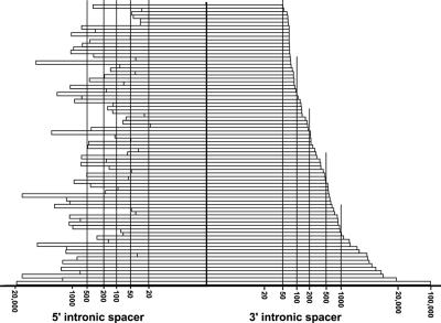
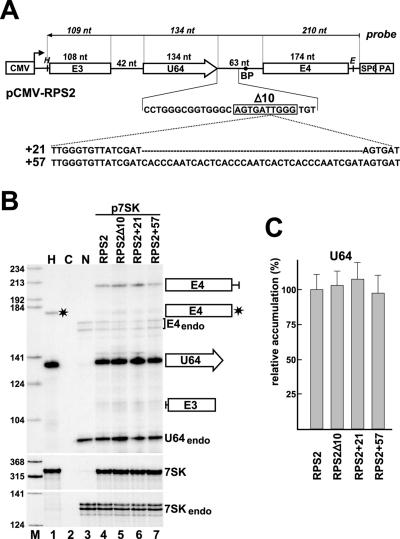
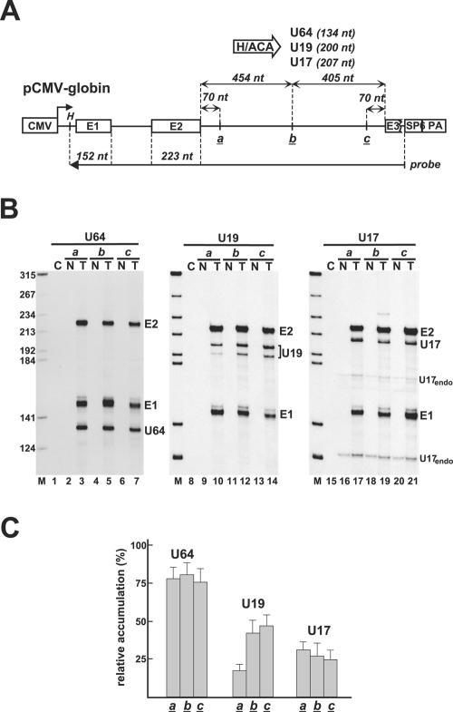
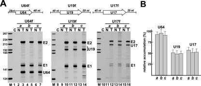
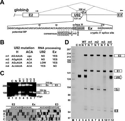
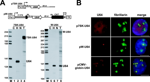
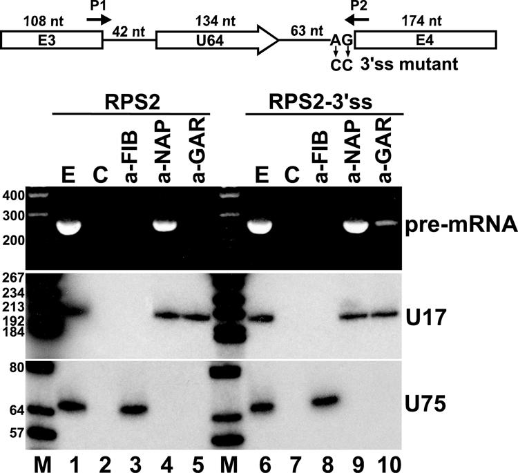
Processing from pre-mRNA introns is a widespread mechanism to generate human box C/D and H/ACA snoRNAs. Recent studies revealed that an optimal position relative to the 3' splice site is important for efficient processing of most box C/D snoRNAs and that assembly of box C/D snoRNPs is stimulated by splicing factors likely bound to the branch point region. Here we have investigated the processing of another major class of human intron-encoded RNAs, the box H/ACA snoRNAs. Analysis of 80 H/ACA RNA genes revealed that human H/ACA RNAs possess no preferential localization close to the 3' or 5' splice site. In vivo processing experiments confirmed that H/ACA intronic snoRNAs are processed in a position-independent manner, indicating that there is no synergy between H/ACA RNA processing and splicing. We also showed that recognition of intronic H/ACA snoRNAs and assembly of pre-snoRNPs is an early event that occurs during transcription elongation parallel with pre-mRNA splice site selection. Finally, we found that efficient processing and correct nucleolar localization of the human U64 H/ACA snoRNA requires RNA polymerase II-mediated synthesis of the U64 precursor. This suggests that polymerase II-associated factors direct the efficient assembly and determine the correct subnuclear trafficking of human H/ACA snoRNPs.


Figures

FIG. 1.
FIG. 1.
Distribution of the length of intronic spacer sequences separating 80 human intron-encoded box H/ACA RNAs from adjacent 5′ and 3′ splice sites. The numbers of upstream and downstream intronic flanking nucleotides are indicated on logarithmic scales.
FIG. 2.
FIG. 2.
In vivo processing of human U64 box H/ACA snoRNA from the third intron of ribosomal protein S2 pre-mRNA. (A) Schematic structure of expression constructs. A fragment of the human ribosomal protein S2 gene encompassing exons 3 and 4 (E3 and E4) and the third intron carrying the U64 gene (open arrow) was fused to the cytomegalovirus promoter (CMV). The polyadenylation region (PA) and the promoter of the SP6 RNA polymerase are indicated. Relevant restriction sites are shown (H, HindIII; E, EcoRI). The antisense RNA probe used for RNase A/T1 mapping and the expected sizes of protected fragments (in italics) are shown. The downstream intronic flanking sequences of U64 as well as sequences deleted from RPS2-Δ10 (boxed) or inserted into the RPS2+21 and RPS2+57 genes are indicated. (B) RNase A/T1 protection. RNAs obtained from mouse L929 transfected with the indicated expression constructs were mapped with a sequence-specific antisense RNA probe, and the protected fragments were separated on a 6% sequencing gel. Control mappings with HeLa (H) and nontransfected L929 cellular RNA (N) and with Escherichia coli tRNA (C) are shown. Positions and structures of protected fragments are indicated on the right. Asterisks indicate a probe fragment protected by the fourth exon of the human HeLa RPS2 mRNA. E4endo, U64endo, and 7SKendo fragments represent endogenous mouse RPS2 exon 4, U64 snoRNA, and 7SK snRNA sequences that partially protect human antisense RNA probes. Lane M, molecular size markers (HaeIII- and TaqI-digested pBR322). (C) Relative accumulation of U64 snoRNA. The intensities of protected probe fragments were quantified by PhosphorImager. The relative levels of U64 were normalized to the transiently expressed 7SK snRNA.
FIG. 3.
FIG. 3.
Processing of U64, U19, and U17 box H/ACA snoRNAs from the second intron of the human β-globin pre-mRNA. (A) Schematic structure of expression constructs. Exons (E1, E2, and E3) of the globin gene and the insertion sites of U64, U19, and U17 snoRNA genes (a, b, and c) are indicated. (B) RNase A/T1 mapping. RNAs from COS7 cells either nontransfected (N) or transfected (T) with the indicated expression vector were mapped with sequence-specific RNA probes. Protected probe fragments representing the spliced exons (E1 and E2) of the transiently expressed human globin mRNA and the excised U64, U19, and U17 snoRNAs are indicated. Fragments U17endo represent endogenous mouse U17 snoRNA partially protecting the human U17 snoRNA probe. (C) Accumulation of U64, U19, and U17 snoRNAs. The relative levels of U64, U19, and U17 were normalized to the levels of exons 1 and 2. For other details, see the legend to Fig. 2.
FIG. 4.
FIG. 4.
Processing of U64, U19, and U17 box H/ACA snoRNAs flanked with natural intronic sequences. (A) RNase A/T1 mapping. The U64, U19, and U17 snoRNAs genes together with their natural flanking sequences (U64f, U19f, and U17f) were inserted into the a, b, and c sites of pCMV-globin. RNAs obtained from COS7 cells nontransfected (N) or transfected (T) with the indicated expression construct were mapped with cRNA probes. (B) Relative accumulation of U64, U19, and U17 snoRNAs. For other details, see the legend to Fig. 3.
FIG. 5.
FIG. 5.
Processing of globin-b pre-mRNA carrying the U92 box H/ACA scaRNA. (A) Schematic structure of the 3′-terminal part of the globin-b-U92 transcript. Dashed lines indicate alternative splicing patterns. Position of exon x (Ex) (boxed) and sequences at the cryptic 5′ and 3′ splice sites are shown. A potential branch point (BP) is underlined, and the box H motif of U92 is shaded. The consensus sequence at human U2-type 3′ splice sites is shown (8). Arrows represent primers used for RT-PCR. Numbering is from the 5′ end of mature U92 RNA or from the transcription initiation site of the human β-globin gene. (B) Mutations introduced into the H or ACA box of U92. Altered nucleotides are shown by lowercase letters, and the expected consequences on U92 excision and Ex exon inclusion are listed. (C) Analysis of RNA processing by RT-PCR. RNAs obtained from mouse L929 cells nontransfected (N) or transfected with the pCMV-globin-b expression construct carrying the wild-type (WT) or mutant (m1 to m4) U92 RNA genes were analyzed by RT-PCR. Lane C represents control RT-PCR performed with Escherichia coli tRNA. The amplified fragments were separated on a 2% agarose gel, cloned, and subjected to sequence analysis. Sequencing gels representing the E2-Ex and Ex-E3 junction regions are shown. (D) RNase A/T1 mapping. RNAs from mouse cells expressing (T) globin-b pre-mRNAs carrying wild-type (WT) or mutant (m1 to m4) U92 RNAs were mapped with sequence-specific RNA probes spanning the entire globin primary transcripts (see Fig. 3A). Control mappings with nontransfected mouse (N) or HeLa (H) cellular RNAs or E. coli tRNA (C) are shown.
FIG. 7.
FIG. 7.
Correct expression of U64 snoRNP depends on pol II transcription. (A) Processing of U64 RNA from pol I and pol III transcripts. Schematic structures of the pol III-specific p7SK-U64 and the pol I-specific pW-U64 expression constructs are shown. The human intronic U64 snoRNA gene, together with 28-bp upstream and 48-bp downstream flanking sequences, was fused to the promoter (Pol III) and terminator (TTTTT) of the human 7SK snRNA gene or inserted into the XbaI (Xb) and XhoI (Xh) sites of the pW ribosomal minigene (24). The mouse pol I promoter (Pol I) and terminator (TR), fragments derived from the 5′ (hatched boxes) and 3′ (open box) external transcribed spacers (ETS) of the mouse rRNA gene, and a fragment of the chloramphenicol acetyltransferase (CAT) gene are shown. RNAs extracted from HeLa (H) cells, transfected (T) or nontransfected (N) COS7 cells (lanes 3 and 4), or mouse L929 (lanes 7 and 8) cells were analyzed by RNase A/T1 mapping with sequence-specific probes as indicated above the lanes. Lane C represents control mapping with Escherichia coli tRNA. (B) In situ localization. COS7 cells transfected with the pCMV-globin-U64 or p7SK-U64 and mouse L929 transfected with the pW-U64 expression constructs were hybridized with a fluorescent oligonucleotide probe specific for human U64. The nucleolus was visualized by staining with an anti-fibrillarin antibody. Nuclear DNA was stained with DAPI (blue). Bar, 10 μm.
FIG. 6.
FIG. 6.
Coimmunoprecipitation of RPS2 pre-mRNA with dyskerin. A schematic structure of transiently expressed human RPS2 pre-mRNA hosting U64 snoRNA is shown. Altered nucleotides in RPS2-3′ss mutant pre-mRNA are shown. Arrows P1 and P2 represent deoxyoligonucleotide primers used for PCRs. For other details, see the legend to Fig. 2A. Cellular extracts prepared from mouse L929 cells transfected with pCMV-RPS2 or pCMV-RPS2-3′ss were treated with protein A-agarose-bound antifibrillarin (a-FIB), antidyskerin (a-NAP), or anti-GAR1 (a-GAR) antibody. RNAs recovered from the extracts (E) and protein A agarose beads either containing or lacking (C) antibodies were analyzed by RT-PCR (upper panel) or Northern blot analysis (lower panels). The amplified RT-PCR fragments were analyzed on 2% agarose gel. For Northern blot analysis, RNAs were separated on a 6% sequencing gel, electrotransferred onto a nylon membrane, and probed with terminally labeled U17- and U75-specific deoxyoligonucleotides. Lanes M represent size markers in base pairs (upper panel) or in nucleotides (lower panels).

Similar articles

Cited by

References

    1. Ambros, V. 2004. The functions of animal microRNAs. Nature 431:350-355. - PubMed
    1. Bachellerie, J. P., J. Cavaillé, and A. Hüttenhofer. 2002. The expanding snoRNA world. Biochimie 84:775-790. - PubMed
    1. Baker, D. L., O. A. Youssef, M. I. Chastkofsky, D. A. Dy, R. M. Terns, and M. P. Terns. 2005. RNA-guided RNA modification: functional organization of the archaeal H/ACA RNP. Genes Dev. 19:1238-1248. - PMC - PubMed
    1. Ballarino, M., M. Morlando, F. Pagano, A. Fatica, and I. Bozzoni. 2005. The cotranscriptional assembly of snoRNPs controls the biosynthesis of H/ACA snoRNAs in Saccharomyces cerevisiae. Mol. Cell. Biol. 25:5396-5403. - PMC - PubMed
    1. Bartel, D. P. 2004. MicroRNAs: genomics, biogenesis, mechanism, and function. Cell 116:281-297. - PubMed

Publication types

MeSH terms

LinkOut - more resources