. 2006 Apr;26(7):2570-82.
doi: 10.1128/MCB.26.7.2570-2582.2006.

Cyclin D3 maintains growth-inhibitory activity of C/EBPalpha by stabilizing C/EBPalpha-cdk2 and C/EBPalpha-Brm complexes

Affiliations

Cyclin D3 maintains growth-inhibitory activity of C/EBPalpha by stabilizing C/EBPalpha-cdk2 and C/EBPalpha-Brm complexes

Guo-Li Wang et al. Mol Cell Biol. 2006 Apr.

Abstract

C/EBPalpha arrests proliferation of young livers by inhibition of cdk2. In old mice, C/EBPalpha inhibits growth by repression of E2F-dependent promoters through the C/EBPalpha-Brm complex. In this paper, we show that cyclin D3-cdk4/cdk6 supports the ability of C/EBPalpha to inhibit liver proliferation in both age groups. Although cyclin D3-cdk4/cdk6 kinases are involved in the promotion of growth, they are expressed in terminally differentiated cells, suggesting that they have additional functions in these settings. We demonstrate that C/EBPalpha represents a target for phosphorylation by cyclin D3-cdk4/cdk6 complexes in differentiated liver cells and in differentiated adipocytes. Cyclin D3-cdk4/cdk6 specifically phosphorylate C/EBPalpha at Ser193 in vitro and in the liver and support growth-inhibitory C/EBPalpha-cdk2 and C/EBPalpha-Brm complexes. We found that cyclin D3 is increased in old livers and activates cdk4/cdk6, resulting in stabilization of the C/EBPalpha-Brm complex. Old livers fail to reduce the activity of cyclin D3-cdk4/cdk6 after partial hepatectomy, leading to high levels of C/EBPalpha-Brm complexes after partial hepatectomy, which correlate with weak proliferation. We examined the role of cyclin D3 in the stabilization of C/EBPalpha-cdk2 and C/EBPalpha-Brm by using 3T3-L1 differentiated cells. In these cells, cyclin D3 is increased during differentiation and phosphorylates C/EBPalpha at Ser193, leading to the formation of growth-inhibitory C/EBPalpha-cdk2 and C/EBPalpha-Brm complexes. The inhibition of cyclin D3 blocks the formation of these complexes. Thus, these studies provide a new function of cyclin D3, which is to support the growth-inhibitory activity of C/EBPalpha.


Figures

FIG. 1.
FIG. 1.
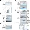
Ser193-phosphorylated isoform of C/EBPα is abundant in C/EBPα-cdk2 and C/EBPα-Brm complexes. A. Characterization of antibodies to the phosphorylated Ser193-C/EBPα isoform. HEK293 cells were transfected with WT and a C/EBPα-S193A mutant, and nuclear extracts were examined by Western blotting with antibodies to ph-Ser193 of C/EBPα and with Abs to total C/EBPα. The filter was reprobed with antibodies to β-actin. B. Western blotting of C/EBPα immunoprecipitates with antibodies to Ser193-ph. C/EBPα was immunoprecipitated from nuclear extracts of young and old livers and examined by Western blotting with Ser193-ph antibodies. The membrane was reprobed with antibodies to total C/EBPα. Immunoglobulin G detected on the same filter showed an equal addition of antibodies to total C/EBPα. C. Phosphorylation of C/EBPα at Ser193 is increased in old livers. Bar graphs show a summary of three experiments with the IP-Western approach. A ratio of signals of ph-Ser193 to total C/EBPα was determined by densitometry. D and E. Examination of C/EBPα isoforms in young and old livers by 2D-Western. The upper image shows a 2D analysis of a control sample of C/EBPα which was overexpressed in cultured cells. Untreated and CIP-treated nuclear proteins were separated by 2D electrophoresis and examined by Western blotting with antibodies to C/EBPα. The percentage of Ser193 isoforms was calculated by densitometry as a ratio to the total amount of C/EBPα isoforms (E). F. Isolation of C/EBPα-cdk2 and C/EBPα-Brm complexes for examination of phosphorylation of C/EBPα. Upper image: nuclear extracts from young livers were fractionated by size exclusion chromatography on an SEC250 column. Identical fractions of five HPLC runs were combined, and positions of C/EBPα, Rb, Brm, and cdk2 within fractions were determined by Western blotting. The location of the C/EBPα-cdk2 complex was determined by co-IP (C/EBPα IP, cdk2 Western). Bottom image: localization of the C/EBPα-Brm complex within fractions of the size exclusion chromatography column with nuclear extracts from old livers. Identical fractions of five runs of old livers on the SEC400 column were combined and examined by Western blotting with antibodies to Brm, cdk2, C/EBPα, and Rb. C/EBPα was immunoprecipitated from each fraction, and Rb was detected in these IPs (C/EBPα-IP, Rb-Western; bottom image). G. 2D analysis of C/EBPα within cdk2 complexes and within the age-specific C/EBPα-Brm complex. C/EBPα complexes were immunoprecipitated from fractions containing the complexes and examined by 2D gel electrophoresis. One half of each IP was treated with alkaline phosphatase (CIP) and examined in a parallel run. Positions of Ser193-phosphorylated isoforms are shown by red arrows.
FIG. 2.
FIG. 2.
cdk4 and cdk6 phosphorylate Ser193 of C/EBPα in vitro and in vivo. A. Cyclin D3-cdk4 and cyclin D3-cdk6 phosphorylate C/EBPα in liver. cdk4, cdk6 and cyclin D3 were immunoprecipitated from young and old livers and examined in a kinase assay with GST-C/EBPα substrate. Ag, agarose control for absorption. The bottom part shows a Coomassie stain of the filter to verify loading of the GST-C/EBPα substrate. B. Baculovirus-expressed, purified cdk4-cyclin D1 phosphorylates C/EBPα. Full-length GST-C/EBPα or GST-140-200 (growth-inhibitory region of C/EBPα) was incubated with the cyclin D1-cdk4 complex in a kinase assay mixture containing [γ-32P]ATP. Proteins were transferred on the membrane and exposed. The bottom image shows Western blotting of the same membrane with antibodies to cdk4. C. C/EBPα forms a stable complex with cyclin D1-cdk4 in the kinase reactions. C/EBPα was incubated with cdk4-cyclin D1 in a kinase reaction, cyclin D1 was immunoprecipitated, and cdk4 and C/EBPα in IPs were examined by Western blotting. A control experiment was performed with bovine serum albumin (BSA) instead of C/EBPα. An aliquot from each reaction mixture was incubated with the GST-Rb substrate in the presence of [γ-32P]ATP (bottom part). D. Baculovirus-expressed, purified cdk4-cyclin D1 and cdk6-cyclin D3 phosphorylate Ser193 of C/EBPα. GST-WT-C/EBPα (W) and a C/EBPα-S193A mutant (S193) were incubated with cdk4-cyclin D1 and with cdk6-cyclin D3 in a kinase assay. Positions of C/EBPα and a nonspecific band (NS for cdk4-cyclin D1) are shown. The filter was reprobed with Abs to C/EBPα, cdk4, and cdk6 (Western). E. cdk4-cyclin D3 phosphorylates Ser193 of C/EBPα. WT and S193A mutant GST-C/EBPα were incubated with cdk4 and cyclin D3 IPs from old livers. The bottom image shows GST-C/EBPα stained with Coomassie. F. cdk6-cyclin D3 phosphorylates Ser193 of C/EBPα. WT and S193A mutant GST C/EBPα were incubated with cdk6 and cyclin D3 IPs from old livers. The bottom image shows GST-C/EBPα stained with Coomassie. G. Examination of efficiency of phosphorylation of C/EBPα by cdk4 relative to phosphorylation of Rb. GST-Rb (0.5 μg) and increasing amounts (0.5, 1, and 2 μg) of the growth-inhibitory region of C/EBPα (GST-C/EBPα-140-200) were added into kinase reaction mixtures with purified cyclin D1-cdk4 and [γ-32P]ATP. The reaction mixtures were loaded on a denaturing PAAG, transferred on membrane, and exposed to X rays. The bottom part shows a Coomassie stain of the gel loaded with the corresponding ratios of the proteins.
FIG. 3.
FIG. 3.
Aging liver increases activities of cdk4 and cdk6 by elevation of cyclin D3. A and B. Protein levels of cyclin D3 are increased in old livers. Nuclear extracts from five livers of young and old animals were examined by Western blotting with antibodies to cyclin D3. The membrane was reprobed with Abs to β-actin. Coomassie stain shows staining of a parallel gel loaded with the same proteins. Bar graphs (B) represent a summary of multiple experiments. Levels of cyclin D3 were calculated as a ratio to β-actin. C. C/EBPα is observed in complexes with cyclin D3 in the liver. Cyclin D3 was immunoprecipitated from young and old livers, and cdk4, cdk6, and C/EBPα were examined by Western blotting in cyclin D3 IPs. IgG lanes, heavy chains of immunoglobulin G were detected by Coomassie staining of the membrane. An aliquot from each reaction mixture was incubated with GST-C/EBPα substrate in the presence of [γ-32P]ATP (kinase assay, bottom part). D. cdk4-cyclin D3 are associated with the C/EBPα-Brm complex in old livers. Nuclear extracts from young and old livers were fractionated by size exclusion chromatography on an SEC-400 column as described in the legend to Fig. 1. Positions of molecular mass markers are shown on the top. Western lanes: examination of cdk4, cdk6, cyclin D3, and C/EBPα in the gel filtration fractions by Western blotting. C/EBPα-IP lanes, C/EBPα was immunoprecipitated from fractions and C/EBPα IPs were probed with antibodies to cyclin D3. Kinase assay lanes: cdk4 and C/EBPα were immunoprecipitated from each fraction and examined in a kinase assay with GST-Rb and with GST-C/EBPα substrates. Positions of cyclin D3-cdk4/cdk6 complexes are shown on the bottom.
FIG. 4.
FIG. 4.
Old livers fail to dephosphorylate C/EBPα at Ser193 after partial hepatectomy and contain a high-MW cyclin D3-cdk4-C/EBPα-Rb-E2F4-Brm complex. A. Expression of cyclin D3 and PP2A in young and old livers at early time points after partial hepatectomy. Nuclear extracts were isolated at 0, 4, and 8 h after PH and examined by Western blotting with antibodies to cyclin D3, cyclin D1, cdk4, cdk6, PP2A, C/EBPα, and C/EBPβ. Each membrane was reprobed with antibodies to β-actin. β-Actin controls are shown for cyclin D3 and C/EBPα membranes. Coomassie stain shows staining of the membrane for cyclin D3. Cyclin D3 IP-kinase assay: examination of cdk4/cdk6 activities in cyclin D3 IPs during early steps of liver proliferation. Cyclin D3 was immunoprecipitated from nuclear extracts isolated at 0, 4, and 8 h after PH. The activities of cdk4 and cdk6 associated with cyclin D3 were examined in a kinase assay with GST-C/EBPα substrate. C/EBPα-IP-Western: C/EBPα was immunoprecipitated from nuclear extracts with antibodies to total C/EBPα. IPs were probed with Abs to cyclin D3 and with Abs to the Ser193-ph isoform of C/EBPα. B. The growth-inhibitory Ser193-ph isoform of C/EBPα is abundant in old livers after PH. C/EBPα was examined by 2D gel electrophoresis in nuclear extracts from young and old livers at 0 and 4 h after PH. Positions of five major isoforms of C/EBPα are shown on the top. Ser193-ph isoforms (a and b) are shown in red. C. Examination of C/EBPα-Brm complex in young and old livers after PH by the co-IP approach. C/EBPα was immunoprecipitated from nuclear extracts isolated from young and old livers at 4 h after PH, and IPs were examined by Western blotting with antibodies to C/EBPα, Brm, Rb, cyclin D3, and cdk4. IgG, heavy chains of immunoglobulin G. The composition of the cyclin D3-C/EBPα-Brm complex is shown on the right. D. Examination of the cyclin D3-C/EBPα-Brm complex in nuclear extracts isolated 4 h after PH by size exclusion chromatography. Nuclear extracts from young and old livers were examined by HPLC-based gel filtration as described in the legend to Fig. 3. C/EBPα-IP lanes: C/EBPα was immunoprecipitated from each fraction, and the presence of cyclin D3 and Brm was examined in these IPs by Western blotting. IgG lanes: heavy chains of immunoglobulin G were determined on the corresponding filters by Coomassie staining.
FIG. 5.
FIG. 5.
Induction of cyclin D3 in differentiated 3T3-L1 cells is required for the phosphorylation of C/EBPα at Ser193. A. Protein levels of cyclin D3 are increased in 3T3-L1 after induction of differentiation. Nuclear extracts were isolated from 3T3-L1 cells at different days after initiation of differentiation (shown on the top) and examined by Western blotting with antibodies to the proteins shown on the left. Protein levels of cyclin D3 were calculated as ratios to β-actin and are shown in the bar graph. B. Expression of markers of 3T3-L1 differentiation, PPARγ and C/EBPβ, and cyclin-dependent kinases. Western blotting was performed with nuclear extracts isolated from 3T3-L1 differentiated cells as described in Materials and Methods. C. Phosphorylation of C/EBPα during differentiation of 3T3-L1 cells. Nuclear extracts from differentiated 3T3-L1 cells were examined by Western blotting with antibodies to total C/EBPα and to the ph-S193 isoform of C/EBPα. D. Examination of activities of kinases associated with cyclin D3. Cyclin D3 was immunoprecipitated from nuclear extracts and added into an in vitro kinase assay mixture with GST-C/EBPα and GST-Rb substrates. The membranes were stained with Coomassie to visualize substrates. E. Inhibition of cyclin D3 expression by siRNA blocks phosphorylation of C/EBPα at Ser193. Control siRNA lanes: expression of cyclin D3 in 3T3-L1 cells which were treated with control (random sequence) siRNA. The filter was reprobed with β-actin to verify protein loading. Cyclin D3 siRNA lanes: siRNA to cyclin D3 was transfected into 3T3-L1 cells 1 day before initiation of differentiation. Proteins extracts were isolated from cyclin D3 siRNA-treated 3T3-Ll cells at different days and examined by Western blotting with antibodies to cyclin D3, cdk4, PPARγ, total C/EBPα, and the Ser193-ph isoform of C/EBPα. Each membrane was reprobed with Abs to β-actin. The bar graph (bottom) presents levels of cyclin D3 in control 3T3-L1 cells (treated with control siRNA) and in 3T3-L1 cells treated with siRNA to cyclin D3 and is a summary of three independent experiments. Cyclin D3 levels were calculated as ratios to β-actin and then as ratios to levels at day 0. Cyclin D3-IP kinase lanes: examination of the cdk4/cdk6 activities in cyclin D3 IPs using GST-C/EBPα as substrate.
FIG. 6.
FIG. 6.
Phosphorylation of C/EBPα by cyclin D3-cdk4/cdk6 is required for the formation of the C/EBPα-cdk2 and the C/EBPα-Brm complexes. A. Examination of C/EBPα-cdk2 and C/EBPα-Brm complexes during differentiation of 3T3-L1 cells treated with control siRNA (upper) and with siRNA to cyclin D3 (bottom). Input lanes: Western blotting of nuclear extracts with antibodies to Brm and cdk2, showing the levels of these proteins in extracts used for the co-IP. C/EBPα-IP lanes: C/EBPα was immunoprecipitated from nuclear extracts, and the IPs were examined by Western blotting with antibodies to cdk2, Brm, and C/EBPα. Heavy chains of immunoglobulin G (IgG) are shown for each IP. B. Examination of the cyclin D3-C/EBPα-Brm complex in differentiated 3T3-L1 cells by size exclusion chromatography. Nuclear extracts from differentiated 3T3-L1 cells (day 7) were fractionated with an SEC-400 column. Fractions were analyzed by Western blotting with antibodies to the proteins shown on the right. C/EBPα-IP Western lanes: examination of cyclin D3 and cdk4 in C/EBPα IPs from size exclusion fractions was performed with specific antibodies as described in the legends to Fig. 3 and 4. Kinase activity of cyclin D3-cdk4 was examined in C/EBPα IPs by an in vitro kinase assay with Rb substrate (C/EBPα-IP kinase). C. Size exclusion chromatography of nuclear extracts isolated from 3T3-L1 cells treated with control siRNA and with cyclin D3 siRNA. Proteins were isolated at day 7 after initiation of differentiation and fractionated on an SEC-400 column. Size exclusion fractions were examined by Western blotting with antibodies to C/EBPα. Amounts of C/EBPα were calculated in fractions containing C/EBPα-Brm complexes relative to total amounts of C/EBPα throughout the gel filtration.
FIG. 7.
FIG. 7.
Hypothetic model suggesting that cyclin D3 supports quiescence of the liver and differentiated adipocytes by stabilizing growth-inhibitory C/EBPα-cdk2 and C/EBPα-Brm complexes. Cyclin D3 activates cdk4/cdk6 kinases, which specifically phosphorylate Ser193 of C/EBPα. The Ser193-ph isoform of C/EBPα forms complexes with cdk2 (young livers) and with Brm, Rb, and E2F4 (old livers), which inhibit cell proliferation.

Similar articles

Cited by

References

    1. Bartkova, J., J. Lukas, M. Strauss, and J. Bartek. 1998. Cyclin D3: requirement for G1/S transition and high abundance in quiescent tissues suggest a dual role in proliferation and differentiation. Oncogene 17:1027-1037. - PubMed
    1. Cenciarelli, C., F. De Santa, P. L. Puri, E. Mattei, L. Ricci, F. Bucci, A. Felsani, and M. Caruso. 1999. Critical role played by cyclin D3 in the MyoD-mediated arrest of cell cycle during myoblasts differentiation. Mol. Cell. Biol. 19:5203-5217. - PMC - PubMed
    1. Conboy, I. M., M. J. Conboy, A. J. Wagers, E. R. Girma, I. L. Weisman, and T. A. Rando. 2005. Rejuvenation of aged progenitor cells by exposure to a young systemic environment. Nature 43:760-764. - PubMed
    1. de La Serna, I. L., K. Roy, K. A. Carlson, and A. N. Imbalzano. 2001. MyoD can induce Cell cycle arrest but not muscle differentiation in the presence of dominant negative SWI/SNF chromatin remodeling enzymes. J. Biol. Chem. 276:41486-41491. - PubMed
    1. Doglioni, C., C. Chiarelli, E. Macri, A. P. Dei Tos, E. Meggiolaro, P. Dalla Palma, and M. Barbaresch. 1998. Cyclin D3 expression in normal, reactive and neoplastic tissues. J. Pathol. 185:159-166. - PubMed

Publication types

MeSH terms