. 2006 Apr;26(7):2583-94.
doi: 10.1128/MCB.26.7.2583-2594.2006.

Role for centromeric heterochromatin and PML nuclear bodies in the cellular response to foreign DNA

Affiliations

Role for centromeric heterochromatin and PML nuclear bodies in the cellular response to foreign DNA

Cleo L Bishop et al. Mol Cell Biol. 2006 Apr.

Erratum in

  • Mol Cell Biol. 2006 May;26(10):3997

Abstract

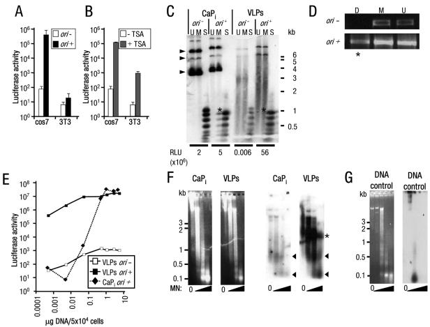
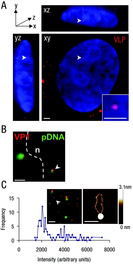
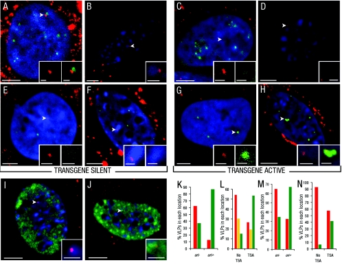
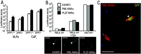
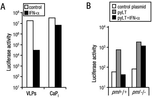
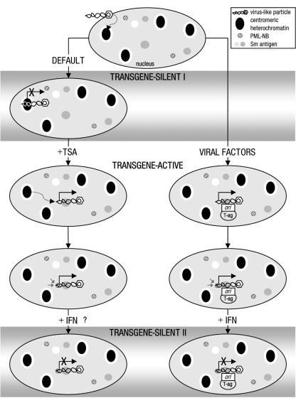
Nuclear spatial positioning plays an important role in the epigenetic regulation of eukaryotic gene expression. Here we show a role for nuclear spatial positioning in regulating episomal transgenes that are delivered by virus-like particles (VLPs). VLPs mediate the delivery of plasmid DNA (pDNA) to cell nuclei but lack viral factors involved in initiating and regulating transcription. By tracking single fluorescently labeled VLPs, coupled with luciferase reporter gene assays, we found that VLPs transported pDNA to cell nuclei efficiently but transgenes were immediately silenced by the cell. An investigation of the nuclear location of fluorescent VLPs revealed that the pDNAs were positioned next to centromeric heterochromatin. The activation of transcription by providing viral factors or inhibiting histone deacetylase activity resulted in the localization to euchromatin regions. Further, the activation of transcription induced the recruitment of PML nuclear bodies (PML-NBs) to the VLPs. This association did not play a role in regulating transgene expression, but PML protein was necessary for the inhibition of transgene expression with alpha interferon (IFN-alpha). These results support a model whereby cells can prevent foreign gene expression at two levels: by positioning transgenes next to centromeric heterochromatin or, if that is overcome, via the type I IFN response facilitated by PML-NB recruitment.


Figures

FIG. 1.
FIG. 1.
High-level, copy number-independent VLP-delivered transgene expression activated by viral factors or TSA. (A and B) Luciferase activity (RLU/mg total protein) from (A) cos7 or 3T3 cells treated with VLPs carrying pDNA without (−) or with (+) ori and from (B) cos7 or 3T3 cells treated with VLPs carrying pDNA without ori, cultured without or with TSA. Note: mock VLP-treated cells cultured with TSA resulted in less than 100 RLU/mg total protein (data not shown); y axis is a log scale. Error bars indicate the standard errors. (C) Southern blot analysis of low-molecular-weight DNA Hirt extracts (26) from cos7 cells transfected using CaPi or treated with VLPs carrying pDNA without (−) or with (+) ori. Restriction endonuclease digests of DNA extracts (U, mock-digested DNA; M, MboI [inhibited by dam methylation, digests only DNA replicated in mammalian cells]; or S, Sau3A [digests all DNA]) demonstrated pDNA replication products present in CaPi-transfected but not VLP-treated cells (indicated by asterisks). The positions of migration of form I, II, and III DNAs are indicated by arrowheads. Considerable degradation is observed for the VLP-delivered pDNA, presumably due to the majority of the plasmid being unprotected from nuclease attack in the cell (52). Luciferase activity (RLU/mg total protein) from an aliquot of each transfected cell culture taken prior to Hirt extraction is given below the appropriate tracks. Positions of migration of molecular weight standard markers are shown to the right of the gel. (D) PCR analyses of low-molecular-weight DNA extracts from cos7 cells treated with VLPs as described for panel C. Plasmid-specific PCR amplification was performed after digestion of the extracts with the restriction endonucleases DpnI (D, inhibited by mammalian methylation; PCR amplification detects mammalian replicated pDNA only; indicated by asterisks) or MboI (M, inhibited by dam methylation; PCR amplification detects bacterial input pDNA only) or mock-digested DNA (U). (E) Luciferase activity from cos7 cells treated with increasing amounts of VLPs or CaPi (equivalent to 0.0005 to 5 μg pDNA/5 × 104 cells) carrying pDNA without (−) or with (+) ori. Note: both axes are a log scale. (F) Micrococcal nuclease assays of HeLa cell nuclei transfected with CaPi or treated with VLPs. Following electrophoresis of micrococcal nuclease (MN)-treated samples, the agarose gels were stained with ethidium bromide (left panels) and then subjected to Southern blot analysis (right panels). Consistent with previous observations (29), atypical nucleosome ladders for CaPi-transfected pDNA were detected, represented by protected pDNA fragments of approximately 0.1 and 0.6 kb (arrowheads). Similar ladders were detected for VLP-delivered pDNA. A further band of VLP-protected pDNA fragments (52) of 1.2 to 1.5 kb was also detected (asterisks). (G) Untransfected HeLa cell nuclei with pDNA added to isolated nuclei prior to micrococcal nuclease digestion and then analyzed as described above for panel F. Left panel, ethidium bromide stain; right panel, Southern blot analysis. Positions of migration of molecular weight standard markers are shown to the left of the gels.
FIG. 2.
FIG. 2.
VLPs enter the nuclei of transgene-silent and transgene-active cells. (A) Three-dimensional reconstruction of sequential confocal sections of a cos7 cell nucleus in xy, xz, and yz planes, showing the sections containing a single nuclear Cy3-VLP (red; arrowhead). Nuclear DNA stained with DAPI (blue). Inset, digital zoom of single nuclear Cy3-VLP. Bars, 2 μm (main panel) and 1 μm (inset). (B) Overlay confocal images of Cy3-VLPs (red) and AlexaFluor-488-labeled pDNA (green), showing a single nuclear Cy3-VLP (arrowhead), nuclear boundary (dotted line), and nucleus (n). Control experiments with unlabeled pDNA gave a low-level diffuse signal corresponding to secondary antibody binding (data not shown). Bar, 2 μm. (C) VLPs labeled with Cy3 (on VP1 nanospheres; red) and AlexaFluor-488 (on pDNA; green) were dropped onto glass, and a single confocal section was captured (inset). Intensities of 100 individual Cy3 spots were measured and plotted according to fluorescence intensities. The intensity peak corresponding to a single VLP (approximately 2,000 arbitrary units as described previously) (23) and the fluorescence signal corresponding to a single Cy3-labeled VP1 nanosphere associated with a single AlexaFluor-488-labeled pDNA is indicated (inset of left panel, white arrowhead). Similar complexes were dropped onto mica and analyzed by AFM (inset right; height ranges are indicated by the bar to the right). Bars, 1 μm (inset left) and 200 nm (inset right).
FIG. 3.
FIG. 3.
Location of VLPs with respect to centromeric heterochromatin, euchromatin regions, and PML-NBs. (A to J) Location of Cy3-VLPs relative to centromeric heterochromatin or euchromatin regions or PML-NBs in cos7 cells treated with Cy3-VLPs carrying pDNA without (transgene silent) (A and E) or with ori (transgene active) (C and G) or in 3T3 cells cultured without (transgene silent) (B and F) or with TSA (transgene active) (D and H). The top panels show representative overlay images of single confocal sections with nuclear Cy3-VLPs (red; arrowheads; determined as described in the legend for Fig. 2A) relative to immunostaining for centromeric proteins in cos7 cells (green) with DAPI-stained nuclei (blue) (A and C) or Hoechst 33342-stained nuclear regions in 3T3 cells (blue) (B and D). The middle panels show representative overlay images of single confocal sections with nuclear Cy3-VLPs (red; arrowheads) relative to immunostaining for PML protein (green) with DAPI-stained nuclei (blue) in cos7 cells (E and G) or Hoechst 33342-stained regions (blue) in 3T3 cells (F and H). Note: the signals for immunostained PML protein in panels E and F are less intense than for those in panels G and H as in the former, PML-NBs are not in the same sections as the VLPs. The bottom panels show representative overlay images of single confocal section with nuclear Cy3-VLPs (red; arrowheads) relative to immunostaining for Sm antigen (green)- and Hoechst 33342-stained nuclear regions (blue) in 3T3 cells cultured without (I) or with (J) TSA. Insets show digital zooms of Cy3-VLPs with corresponding cellular structure. (K through N) Graphical representation of the percentage of VLPs in each nuclear location in cos7 cells without (−) or with (+) ori or 3T3 cells without or with TSA, with respect to location next to centromeric heterochromatin or euchromatin regions (red and green bars, respectively, in panel K); centromeric heterochromatin, euchromatin regions, or intermediate regions (red, green or yellow bars, respectively, in panel L); or away from or next to PML-NBs (red and green bars, respectively, in panels M and N). Using chi-squared analysis (described in Material and Methods) the observed difference in Cy3-VLP nuclear location following treatment with TSA or delivery of ori for each panel was statistically significant (K through N) (P < 0.001). Bars, 5 μm (main panels) and 1 μm (insets).
FIG. 4.
FIG. 4.
Disruption of PML-NBs does not affect VLP-delivered transgene expression status. (A) Luciferase activity from primary embryo fibroblasts isolated from wild-type (pml+/+) or pml/ mice that were treated with VLPs or transfected using calcium phosphate precipitate (CaPi) cultured without (open bars) or with TSA (closed bars). Error bars indicate the standard errors. (B) Top, Luciferase activity from cells (as indicated) pretransfected with PML-NB-disrupting gene E4ORF3 or PML-RARα or with nondisrupting control gene PLZF-RARα. Cells were cultured without or with TSA as indicated or in the presence of viral factors (cos7 ori+). Bottom, PML-NB disruption was confirmed by immunofluorescence for PML protein (transfected cells are indicated with arrowheads), and overexpression of RARα fusion proteins was confirmed by immunofluorescence for RARα (data not shown). Bar, 10 μm. (C) Fluorescence image of HeLa cells pretransfected with PML-RARα and then treated with VLPs carrying pDNA encoding the EGFP protein and cultured with TSA. A cell overexpressing PML-RARα (red; detected by immunostaining for RARα) and expressing EGFP (green) is indicated by the arrowhead. Cell nuclei were stained with DAPI (blue). Bar, 20 μm.
FIG. 5.
FIG. 5.
PML protein is necessary for inhibition of VLP-mediated expression by IFN-α. (A) Luciferase activity from cos7 cells that were treated with VLPs or transfected using CaPi carrying pDNA with ori cultured without or with IFN-α. (B) Luciferase activity from primary embryo fibroblasts isolated from wild-type (pml+/+) or pml/ mice that were pretransfected with a control plasmid (pEGFP; open bars) or pCMVLT2 carrying the polyomavirus large T antigen (pyLT) gene and then treated with VLPs carrying pDNA with the polyomavirus ori cultured without (open and gray bars) or with IFN-α (closed bars). Data are representative examples of at least three independent experiments.
FIG. 6.
FIG. 6.
Model for a two-phase cellular defense against VLP-delivered pDNA. Hypothetical stages in the response of cells to invasion by foreign genes are given. The sequence of events is derived from the data presented, with the exception of the order of events for activation and repositioning of transgenes in the presence of TSA, since it is not known which occurs first. The locations and relative positions of VLP and cellular components shown are schematic representations derived from fluorescence data. Transgene expression status is based on luciferase assay data. Inhibition of HDAC activity can attenuate the IFN antiviral pathway (44); therefore, the effect of IFN-α treatment on TSA-mediated transgene activation has not been determined.

Similar articles

Cited by

References

    1. An, K., A. Q. Paulsen, M. B. Tilley, and R. A. Consigli. 2000. Use of electron microscopic and immunogold labeling techniques to determine polyomavirus recombinant VP1 capsid-like particles entry into mouse 3T6 cell nucleus. J. Virol. Methods 90:91-97. - PubMed
    1. Baxter, J., M. Merkenschlager, and A. G. Fisher. 2002. Nuclear organisation and gene expression. Curr. Opin. Cell Biol. 14:372-376. - PubMed
    1. Belmont, A. S., S. Dietzel, A. C. Nye, Y. G. Strukov, and T Tumbar. 1999. Large-scale chromatin structure and function. Curr. Opin. Cell Biol. 11:307-311. - PubMed
    1. Boisvert, F. M., M. J. Hendzel, and D. P. Bazett-Jones. 2000. Promyelocytic leukemia (PML) nuclear bodies are protein structures that do not accumulate RNA. J. Cell Biol. 148:283-292. - PMC - PubMed
    1. Boisvert, F. M., M. J. Kruhlak, A. K. Box, M. J. Hendzel, and D. P. Bazett-Jones. 2001. The transcription coactivator CBP is a dynamic component of the promyelocytic leukemia nuclear body. J. Cell Biol. 152:1099-1106. - PMC - PubMed

Publication types

MeSH terms