. 2006 Apr;26(7):2661-74.
doi: 10.1128/MCB.26.7.2661-2674.2006.

Increased DNA damage sensitivity and apoptosis in cells lacking the Snf5/Ini1 subunit of the SWI/SNF chromatin remodeling complex

Affiliations

Increased DNA damage sensitivity and apoptosis in cells lacking the Snf5/Ini1 subunit of the SWI/SNF chromatin remodeling complex

Agnes Klochendler-Yeivin et al. Mol Cell Biol. 2006 Apr.

Abstract

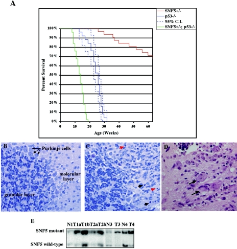
The gene encoding the SNF5/Ini1 core subunit of the SWI/SNF chromatin remodeling complex is a tumor suppressor in humans and mice, with an essential role in early embryonic development. To investigate further the function of this gene, we have generated a Cre/lox-conditional mouse line. We demonstrate that Snf5 deletion in primary fibroblasts impairs cell proliferation and survival without the expected derepression of most retinoblastoma protein-controlled, E2F-responsive genes. Furthermore, Snf5-deficient cells are hypersensitive to genotoxic stress, display increased aberrant mitotic features, and accumulate phosphorylated p53, leading to elevated expression of a specific subset of p53 target genes, suggesting a role for Snf5 in the DNA damage response. p53 inactivation does not rescue the proliferation defect caused by Snf5 deficiency but reduces apoptosis and strongly accelerates tumor formation in Snf5-heterozygous mice.


Figures

FIG. 1.
FIG. 1.
Proliferative defects and cell death in Snf5-depleted MEFs. (A) Schematic representation of the wild-type (Snf5+), floxed (Snf5L), and deleted (Snf5D) alleles of Snf5. Exons 1 to 3 are indicated by boxes and loxP sites by filled triangles. Relevant restriction sites (EcoRI [E], EcoRV [EV]) and the position of the probe are indicated. Cre-mediated recombination eliminates exons 1 and 2. Southern blot (B) and Western blot (C) analyses of DNA and lysates prepared from mock- and AdCre-infected MEFs with the indicated genotypes, 4 and 6 dpi. For Southern blot analysis, the DNA was digested with EcoRI and EcoRV and the Snf5 locus was identified by hybridization with the probe indicated in panel A. (D) Exit of Snf5-deficient MEFs from the cell cycle. Cells of indicated genotypes are shown 6 days after infection (magnification, ×100). (E) Representative growth curves of mock- and AdCre-infected MEFs with the indicated genotypes. The cells were placed in culture at equal densities, infected, counted, and passaged every 2 days at the same density. Experiments were performed on two different MEF cultures of each genotype, and duplicate plates were counted. The curves represent the means of these values.
FIG. 2.
FIG. 2.
Snf5 depletion impairs cell cycle progression and increases the rate of apoptosis under growth-inhibiting conditions. (A) Cell cycle analysis of mock- and AdCre-infected Snf5lox/+ and Snf5lox/ MEFs, 6 days after infection. The cells were cultured in 10% serum-containing medium or serum starved for 72 h before the analysis. DNA content was determined by propidium iodide staining and flow cytometric analysis. (B) BrdU incorporation assays of Snf5lox/+, Snf5lox/, and Snf5lox/lox MEFs that were mock or AdCre infected. The cells were incubated with BrdU for 30 min and processed for immunostaining. The graph is representative of three independent experiments performed on different preparations of MEFs.
FIG. 3.
FIG. 3.
Expression analysis of E2F target genes and induction of p53 in Snf5-deficient cells. Semiquantitative RT-PCR analysis of E2F-responsive genes (A) and p53 target genes (C). Total RNA was isolated from mock (−)- and AdCre (+)-infected Snf5lox/lox, Snf5lox/, and Snf5lox/+ MEFs, 6 days postinfection. PCR was carried out under linear amplification conditions with 0.01 μl and 0.03 μl of the 25-μl cDNA sample. One representative experiment of four is shown. Similar results were obtained with AdCre-infected Snf5lox/lox and Snf5lox/ MEFs (C). GAPDH expression is shown as a control. (B) Western blot analysis of lysates prepared from mock- or AdCre-infected Snf5lox/ and Snf5lox/+ MEFs, 4 and 6 days after infection. The cells were grown in 10% serum containing medium or in 0.1% serum (low serum) for 72 h as indicated.
FIG. 4.
FIG. 4.
p19ARF induction and genotoxic stress in Snf5-depleted cells. (A, C) Semiquantitative RT-PCR analysis of Snf5, p19ARF (A), and p16-INK4A (C). Total RNA was isolated from mock (−)- and AdCre (+)-infected Snf5lox/lox and Snf5lox/+ MEFs, 4 and 6 days after infection as indicated (A) and from Snf5lox/lox, Snf5lox/, and Snf5lox/+ MEFs 6 days after infection (C) and assayed as described in the legend to Fig. 3. GAPDH expression is shown as a control (A). The GAPDH control for panel C is included in Fig. 3A. (B) Protein levels of p19ARF in mock- and AdCre-infected Snf5lox/ and Snf5lox/+ MEFs, 4 and 6 days after infection, were determined by Western blot analysis. The cells were grown in 10% serum-containing medium or in 0.1% serum (low serum) for 72 h as indicated. (D) Phosphorylation of p53 at serine 18 is detected in Snf5-depleted MEFs. Lysates from mock- or AdCre-infected Snf5lox/lox and Snf5lox/+ MEFs were analyzed by Western blot analysis. Extracts from cells harvested 16 h after UV irradiation (10 J/m2) were included as a positive control.
FIG.5.
FIG.5.
Snf5-null cells display increased sensitivity to genotoxic agents, abnormal mitotic features, and amplification of centrosomes. Mock- and AdCre-infected MEFs with the indicated genotypes were plated in six-well culture dishes and exposed to increasing doses of UV light (A) or doxorubicin (B) at day 3 after infection. The percentage of cell death in each well was assessed by trypan blue exclusion 72 h (A) or 24 h (B) after treatment. Each data point represents the mean of six observations. Similar results were obtained in three independent experiments. The difference in UV hypersensitivity between AdCre-infected Snf5lox/lox and Snf5lox/ MEFs is apparently due to experimental variability. All the Snf5-deficient clones (Cre-infected Snf5lox/lox or Snf5lox/) tested were three- to sixfold more sensitive to UV radiation than their heterozygous counterparts, independently of whether the infected cells carried two floxed alleles or one floxed and one null allele. (C, D) Snf5lox/ and SNF5lox/+ MEFs were infected with AdCre and immunostained 4 days after infection. (C) AdCre-infected Snf5lox/ MEFs were stained with anti-α-tubulin (green) and counterstained with DAPI. White arrows indicate micronuclei, and yellow arrows point to interphase bilobed nuclei with bridging chromatin. (D) AdCre-infected Snf5lox/ MEFs were stained for centrosomes with anti-γ-tubulin (green) and counterstained with DAPI. Micronuclei (C) and centrosomes (D) of infected Snf5lox/ MEFs and Snf5lox/+ MEFs were counted for 100 cells per sample in three independent experiments.
FIG. 6.
FIG. 6.
p53-null background does not rescue the slowed proliferation phenotype in Snf5-depleted cells but reduces apoptosis. (A) Growth curves of mock- and AdCre-infected p53-null MEFs with the indicated genotype for the Snf5 locus. The experiment was performed as described for Fig. 1E, and the graph shows a representative of two independent experiments. Western blot (B) and Southern blot (C) analyses of DNA and lysates prepared from mock- and AdCre-infected p53-null MEFs with the indicated genotypes, 5, 12, and 15 days postinfection. (D) p53 wild-type and p53-null MEFs with the indicated genotypes at the Snf5 locus were infected with AdCre and grown in 10% or 0.1% serum-containing medium for 72 h. The percentage of cell death was determined by trypan blue exclusion in three independent experiments. The percentages of cells with a sub-G1 content of DNA as determined by FACS analysis were similar. (E) Semiquantitative RT-PCR analysis of Puma. Total RNA was isolated from mock (−)- and AdCre (+)-infected MEFs with the indicated genotype 6 days after infection and assayed as described in the legend to Fig. 3. GAPDH expression is shown as a control.
FIG. 7.
FIG. 7.
Snf5 and p53 deficiency cooperate in tumorigenesis in vivo. (A) Kaplan-Meier tumor-free survival curves of Snf5+/ (n = 32), p53−/− (n = 25), and Snf5+/; p53−/− (n = 49) mice (confidence interval [C.I.], 95%). The curves were compared pairwise by the log rank test (P < 0.0001). The mean age of survival was 12.5 weeks for Snf5+/; p53−/− mice and 26 weeks for p53−/− mice. At 60 weeks, 72% of Snf5+/ mice were still alive and tumor free. (B to D) Photomicrographs of hematoxylin- and eosin-stained slides from Snf5+/; p53−/− mice (magnification, ×400). Section of normal cerebellum (B) versus section showing infiltration of malignant cells into the molecular layer (C). Black and red arrows point to mitotic and apoptotic figures, respectively. (D) Representative section of a brain rhabdoid tumor. The arrows point to rhabdoid cells with typical prominent nucleoli and eosinophilic hyaline cytoplasmic inclusions. (E) LOH at the Snf5 locus is observed in tumors (T) from Snf5+/; p53−/− mice. Southern blot analysis of DNA extracted from tumors shows that the wild-type Snf5 allele is greatly reduced, while the ratio of mutant to wild-type alleles is roughly 1 in adjacent normal tissues (N).

References

    1. Aloyz, R. S., S. X. Bamji, C. D. Pozniak, J. G. Toma, J. Atwal, D. R. Kaplan, and F. D. Miller. 1998. p53 is essential for developmental neuron death as regulated by the TrkA and p75 neurotrophin receptors. J. Cell Biol. 143:1691-1703. - PMC - PubMed
    1. Ameyar-Zazoua, M., M. B. Wisniewska, L. Bakiri, E. F. Wagner, M. Yaniv, and J. B. Weitzman. 2005. AP-1 dimers regulate transcription of the p14/p19ARF tumor suppressor gene. Oncogene 24:2298-2306. - PubMed
    1. Andreassen, P. R., F. B. Lacroix, O. D. Lohez, and R. L. Margolis. 2001. Neither p21WAF1 nor 14-3-3sigma prevents G2 progression to mitotic catastrophe in human colon carcinoma cells after DNA damage, but p21WAF1 induces stable G1 arrest in resulting tetraploid cells. Cancer Res. 61:7660-7668. - PubMed
    1. Banin, S., L. Moyal, S. Shieh, Y. Taya, C. W. Anderson, L. Chessa, N. I. Smorodinsky, C. Prives, Y. Reiss, Y. Shiloh, and Y. Ziv. 1998. Enhanced phosphorylation of p53 by ATM in response to DNA damage. Science 281:1674-1677. - PubMed
    1. Bates, S., A. C. Phillips, P. A. Clark, F. Stott, G. Peters, R. L. Ludwig, and K. H. Vousden. 1998. p14ARF links the tumour suppressors RB and p53. Nature 395:124-125. - PubMed

Publication types

MeSH terms

Substances

LinkOut - more resources