. 2006 Apr;26(7):2675-87.
doi: 10.1128/MCB.26.7.2675-2687.2006.

Insulin-like growth factor 1 receptor signaling regulates skin development and inhibits skin keratinocyte differentiation

Affiliations

Insulin-like growth factor 1 receptor signaling regulates skin development and inhibits skin keratinocyte differentiation

Marianna Sadagurski et al. Mol Cell Biol. 2006 Apr.

Abstract

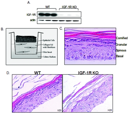
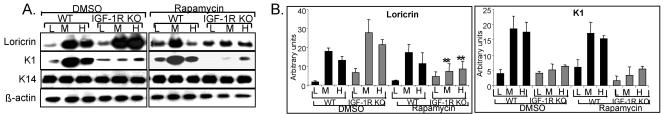
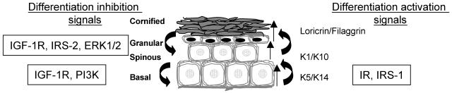
The insulin-like growth factor 1 receptor (IGF-1R) is a multifunctional receptor that mediates signals for cell proliferation, differentiation, and survival. Genetic experiments showed that IGF-1R inactivation in skin results in a disrupted epidermis. However, because IGF-1R-null mice die at birth, it is difficult to study the effects of IGF-1R on skin. By using a combined approach of conditional gene ablation and a three-dimensional organotypic model, we demonstrate that IGF-1R-deficient skin cocultures show abnormal maturation and differentiation patterns. Furthermore, IGF-1R-null keratinocytes exhibit accelerated differentiation and decreased proliferation. Investigating the signaling pathway downstream of IGF-1R reveals that insulin receptor substrate 2 (IRS-2) overexpression compensates for the lack of IGF-1R, whereas IRS-1 overexpression does not. We also demonstrate that phosphatidylinositol 3-kinase and extracellular signal-regulated kinase 1 and 2 are involved in the regulation of skin keratinocyte differentiation and take some part in mediating the inhibitory signal of IGF-1R on differentiation. In addition, we show that mammalian target of rapamycin plays a specific role in mediating IGF-1R impedance of action on keratinocyte differentiation. In conclusion, these results reveal that IGF-1R plays an inhibitory role in the regulation of skin development and differentiation.


Figures

FIG. 1.
FIG. 1.
Lack of IGF-1R expression disturbs skin formation and organization in a 3D organotypic coculture model. (A) Keratinocytes isolated from mice containing the floxed allele of IGF-1R (IGF-1R flox/flox) were maintained in culture for 4 days until reaching confluence. Afterwards, cells were infected with an adenovirus-Cre construct as detailed in Materials and Methods (IGF-1R KO). After 1 h, the viral supernatant was replaced with fresh culture medium. Control cells were infected with an empty viral vector (WT). Total protein lysates were analyzed by Western blotting, using antibodies against the murine IGF-1R. Actin levels are shown, demonstrating equal loading. Evaluation of IGF-1R expression was carried out routinely in all experiments to confirm IGF-1R inactivation. (B) 3D organotypic skin cocultures were established as detailed in Materials and Methods, leading to the formation of a skin-like structure, as depicted in the scheme. (C) Cocultures were maintained for 14 days, after which they were fixed, paraffin embedded, sectioned, and stained with hematoxylin and eosin (H&E). A typical H&E-stained section of the 3D coculture is shown at high (40×) magnification. Layers of epidermal compartments, as occur in vivo, are shown. (D) Skin fibroblasts and keratinocytes were isolated from mice containing the floxed allele of IGF-1R (IGF-1R flox/flox) and infected with the adenovirus-Cre construct as detailed in Materials and Methods. Cells infected with the empty viral vector served as controls (WT). 3D organotypic skin cocultures were established, maintained, and processed as detailed in Materials and Methods. A representative H&E-stained section is shown at high (20×) magnification.
FIG. 2.
FIG. 2.
Decreased proliferation and increased apoptotic rate of the IGF-1R-null organotypic skin coculture. IGF-1R-null (IGF-1R KO) and control (WT) 3D organotypic skin cocultures were established and maintained as detailed in Materials and Methods. (A) Positive PCNA-stained nuclei were detected by immunohistochemistry as described in the text. Photographs of high (40×) magnification are presented. (B) TUNEL staining was detected by immunofluorescence (green [FITC], apoptotic cells; blue [4′,6′-diamidino-2-phenylindole], nuclear counterstaining). For the positive control, normal mice were treated with UVB light and sacrificed 48 h later (b). (a) Nonirradiated mice. (c) Organotypic cocultures of the control (WT); (d) IGF-1R-null cocultures (IGF-1R KO). Percent cell death, determined by tabulating FITC-stained cells, appears in the lower corner (P < 0.05 for WT versus IGF-1R KO). The images demonstrate only the epidermal layer and are representative of results reproduced three to five times.
FIG. 3.
FIG. 3.
Abnormal differentiation pattern of the IGF-1R-null organotypic skin coculture. IGF-1R-null (IGF-1R KO) and control (WT) 3D organotypic skin cocultures were established and maintained as detailed in Materials and Methods. (A) Cocultures were maintained for 14 days, after which they were fixed, paraffin embedded, sectioned, and stained with hematoxylin (upper panels) or stained to demonstrate cell nuclei (lower panels). The ×20 magnification photographs are from a representative experiment. (B) K14, K1, loricrin, and K6 were detected by immunohistochemistry. The high-magnification (×40) photographs are from a representative experiment.
FIG. 4.
FIG. 4.
Lack of IGF-1R expression is associated with decreased proliferation and increased apoptotic rate of primary keratinocytes in vitro. Proliferating IGF-1R flox/flox keratinocytes were grown and maintained as described in the legend for Fig. 1. IGF-1R inactivation was carried as described (IGF-1R KO). Control cells were infected with an empty viral vector (WT). (A) On the day of infection, 24, 48, and 72 h postinfection, live cells were counted; dead cells were excluded by trypan blue staining. (B) The incorporation of BrdU into dividing cells was detected with anti-BrdU antibody as described in Materials and Methods. Cells incorporating BrdU were detected by immunochemistry, as their nucleus acquired a brown color. Photographs of representative fields are shown. Original magnification, ×20. Positively stained nuclei are marked with arrows. (C) The percentage of positively BrdU-stained cells per total number of cells was counted in several fields, arbitrarily chosen, of the WT and IGF-1R KO cultures. *, P < 0.05 for WT versus IGF-1R KO. (D) Primary cultured WT and IGF-1R KO keratinocytes were cultured for 48 h. Apoptotic cells were detected by FACS analysis as described in Materials and Methods using annexin V. The annexin V-positive cell fraction is presented. *, P < 0.05 for WT versus IGF-1R KO. The plot represents an average of three separate experiments.
FIG. 5.
FIG. 5.
Lack of IGF-1R expression facilitates the differentiation process of primary keratinocytes in vitro. Proliferating IGF-1R flox/flox keratinocytes were grown and maintained as described in the legend for Fig. 1. IGF-1R inactivation was carried as described there. Cells were induced to differentiate by increasing the Ca2+ concentration from 0.05 mM (low [L]) to 0.12 mM (medium [M]), or 1.0 mM (high [H]). Cells were further maintained for 12 (A and B) or 48 (C and D) h. The induction of differentiation markers was identified by Western blot analysis, using antibodies against K14, K1, and loricrin. Actin levels are shown and demonstrate equal loading. Results are representative of six independent experiments. Samples of each independent experiment were run on the same gel and blotted together. The blots of loricrin and K1 were further analyzed by scanning densitometry (B and D). Results are a summary (means ± standard errors) of three separate experiments and are expressed in arbitrary units. *, P < 0.01; **, P < 0.001 for WT versus IGF-1R KO, 48 h.
FIG. 6.
FIG. 6.
Differential effects of IRS-1 and IRS-2 proteins on the differentiation process in IGF-1R-null cells. (A) Cells were infected with an adenoviral IRS-1 or IRS-2 construct. Control cells were infected with a luciferase-containing adenoviral construct (Luc). Cell lysates were analyzed by Western blotting with anti-IRS-1 and anti-IRS-2 antibodies, as indicated. IGF-1R inactivation (IGF-1R KO) was carried out as described in the legend for Fig. 1. Control cells were infected with an empty viral vector (WT); at 16 h postinfection, keratinocytes were infected a second time, with an adenoviral IRS-1 (B) or IRS-2 (D) construct. Control cells were infected on both occasions with a luciferase-containing adenoviral construct (Luc); 6 h after the second infection, the cells were induced to differentiate for 48 h as described in the legend for Fig. 5, and analysis of differentiation marker expression was carried out as described there. Actin levels are shown, demonstrating equal loading. Results are representative of three independent experiments. Samples of each independent experiment were run on the same gel and blotted together. The blots of loricrin and K1 were further analyzed by scanning densitometry (C and E). Results are a summary (means ± standard errors) of three separate experiments and are expressed in arbitrary units. **, P < 0.01 for IGF-1R KO luciferase infected versus IGF-1R KO infected by adenoviral IRS-2 construct.
FIG. 7.
FIG. 7.
The PI3K/Akt signaling pathway inhibits skin keratinocyte differentiation. Cells (A and B) were stimulated with IGF-1 (10−7 M) for 3 min. Cells lysates were immunoprecipitated with antiphosphotyrosine antibody and used for an in vitro PI3 kinase assay (A) or were analyzed by Western blotting with anti-phospho-Akt and anti-Akt antibodies (B). Results are representative of three independent experiments. PIP, inositol triphosphate; ORI, origin. (C) Proliferating keratinocytes were infected with DN PI3K, CA PI3K, or luciferase as a control vector (Luc). Thereafter, cells were induced to differentiate for 48 h as already described and analyzed by Western blotting with anti-phospho-Akt and anti-Akt antibodies. (D and F) IGF-1R inactivation was carried as described in the legend for Fig. 1. Proliferating keratinocytes were then infected with DN PI3K (D), CA PI3K (F), or luciferase (Luc) constructs. Cells were induced to differentiate for 48 h as detailed above. Analysis of differentiation marker expression was carried as described in the legend for Fig. 5. Actin levels are shown and demonstrate equal loading. Results are representative of three independent experiments. Samples of each independent experiment were run on the same gel and blotted together. The blots of loricrin and K1 were further analyzed by scanning densitometry (E and G). Results are a summary (means ± standard errors) of three separate experiments and are expressed in arbitrary units. Symbols in panel E: **, P < 0.01 for WT (L and M) luciferase infected versus WT infected by adenoviral DN PI3K construct; *, P < 0.05 for WT (H) luciferase infected versus WT (H) infected by adenoviral DN PI3K construct. Symbols in panel G: **, P < 0.001 for WT (M and H) luciferase infected versus WT infected by adenoviral CA PI3K construct and IGF-1R KO (H) luciferase infected versus IGF-1R KO (H) infected by adenoviral CA PI3K construct.
FIG. 8.
FIG. 8.
Inhibitory role of the MAPK pathway during the differentiation process of skin keratinocytes. Proliferating skin keratinocytes were stimulated with IGF-1 (10−7 M) for 3 min. Cells lysates were immunoprecipitated with anti-phospho myelin basic protein (MBP) antibody and used in an in vitro MAPK activity assay (A) or analyzed by Western blotting with anti-phospho-ERK1/2 or anti-ERK1/2 antibodies (B). (C) IGF-1R inactivation was carried as described in the legend for Fig. 1. Cells were then treated with U0126 (20 μM) and induced to differentiate for 48 h as already detailed. Analysis of differentiation marker expression was carried out as described in the legend for Fig. 5. Actin levels are shown and demonstrate equal loading. Results are representative of three independent experiments. Samples of each independent experiment were run on the same gel and blotted together. The blots of loricrin and K1 were further analyzed by scanning densitometry (D). Results are a summary (means ± standard errors) of three separate experiments and are expressed in arbitrary units. **, P < 0.02 for WT (H) DMSO treated versus WT (H) treated with U0126; *, P < 0.05 for WT (M) DMSO treated versus WT (M) treated with U0126; *, P < 0.05 for IGF-1R KO DMSO treated (M and H) versus IGF-1R KO (M and H) treated with U0126.
FIG. 9.
FIG. 9.
mTOR is a specific mediator of IGF-1R action during keratinocyte differentiation. (A) IGF-1R inactivation was carried out as described as described in the legend for Fig. 1. Thereafter, cells were treated with rapamycin (20 nM) and induced to differentiate for 48 h as detailed in the text. Analysis of differentiation marker expression was carried as described in the legend for Fig. 5. Actin levels are shown and demonstrate equal loading. Samples of each independent experiment were run on the same gel and blotted together. The blots of loricrin and K1 were further analyzed by scanning densitometry (B). Results are a summary (means ± standard errors) of three separate experiments and are expressed in arbitrary units. **, P < 0.01 for IGF-1R KO (M and H) DMSO treated versus (M and H) IGF-1R KO treated with rapamycin.
FIG. 10.
FIG. 10.
Schematic model of the roles of IGF-1R and downstream molecules in epidermal differentiation.

Similar articles

Cited by

References

    1. Accili, D., J. Nakae, J. J. Kim, B. C. Park, and K. I. Rother. 1999. Targeted gene mutations define the roles of insulin and IGF-I receptors in mouse embryonic development. J. Pediatr. Endocrinol. Metab. 12:475-485. - PubMed
    1. Baker, J., J. P. Liu, E. J. Robertson, and A. Efstratiadis. 1993. Role of insulin-like growth factors in embryonic and postnatal growth. Cell 75:73-82. - PubMed
    1. Baserga, R., A. Hongo, M. Rubini, M. Prisco, and B. Valentinis. 1997. The IGF-I receptor in cell growth, transformation and apoptosis. Biochim. Biophys. Acta 1332:F105-F126. - PubMed
    1. Calautti, E., J. Li, S. Saoncella, J. L. Brissette, and P. F. Goetinck. 2005. Phosphoinositide 3-kinase signaling to AKT promotes keratinocyte differentiation versus death. J. Biol. Chem. 280:32856-32865. - PubMed
    1. De Meyts, P., J. Palsgaard, W. Sajid, A. M. Theede, and H. Aladdin. 2004. Structural biology of insulin and IGF-1 receptors. Novartis Found. Symp. 262:160-171. - PubMed

Publication types

MeSH terms