. 2006 Mar 23;49(6):1867-73.
doi: 10.1021/jm051273d.

Enantiomer discrimination illustrated by the high resolution crystal structures of type 4 phosphodiesterase

Affiliations

Enantiomer discrimination illustrated by the high resolution crystal structures of type 4 phosphodiesterase

Qing Huai et al. J Med Chem. .

Abstract

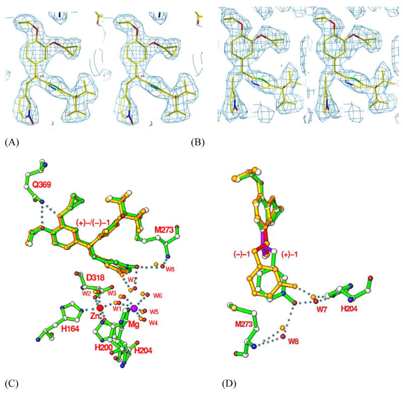
Type 4 phosphodiesterase (PDE4) inhibitors are emerging as new treatments for a number of disorders including asthma and chronic obstructive pulmonary disease. Here we report the biochemical characterization on the second generation inhibitor (+)-1 (L-, IC50=0.4 nM) and its enantiomer (-)-1 (L-, IC50=43 nM) and their cocrystal structures with PDE4D at 2.0 A resolution. Despite the 107-fold affinity difference, both enantiomers interact with the same sets of residues in the rigid active site. The weaker (-)-1 adopts an unfavorable conformation to preserve the pivotal interactions between the Mg-bound waters and the N-oxide of pyridine. These structures support a model in which inhibitors are anchored by the invariant glutamine at one end and the metal-pocket residues at another end. This model provides explanations for most of the observed structure-activity relationship and the metal ion dependency of the catechol-ether based inhibitors and should facilitate their further design.


Figures

Fig. 1
Fig. 1
Chemical structures of PDE4 selective inhibitors. (+)-1 or ()-1 has the chiral center at carbon-9, and can be divided into five subgroups (CORE and R1 to R4) to facilitate discussion.
Fig. 2
Fig. 2
Kinetic properties of (+)-1. (A) Inhibition of PDE4A by (+)-1 with respect to increased cAMP concentration. The linear response has an intercept of 0.43 ± 0.07 nM (apparent Ki) and a slope of 0.19 ± 0.02 nM/μM cAMP. (B). Rapidly reversible PDE4A inhibition by (+)-1. The activity (mean ± se, n = 3) of PDE4A inhibited with 15 nM (+)-1 was determined at 30, 60, 120, and 180 seconds following a 120-fold volume dilution. 100% activity represents the average of the corresponding controls (with DMSO as vehicle). Recovery time represents the dilution time plus the assay duration (60s) except for the 0s point for (+)-1, which represents the enzyme activity in the presence of 15 nM (+)-1.
Fig. 3
Fig. 3
PDE4D2-inhibitor structures. (A) Ribbon diagram of monomeric PDE4D2 catalytic domain. (+)-1 and (+)-1 are shown as the green and golden sticks, respectively. Divalent metal Zn is drawn as a red ball and Mg as a purple ball. (B) Binding of (+)-1 (green sticks and balls) and (+)-1 (golden sticks) at the active site of PDE4D2. The residues of PDE4D2, which are involved in binding with the inhibitors, are labeled. The four metal binding residues are shown in light blue.
Fig. 4
Fig. 4
Binding of (+)-1 and ()-1 (L869298 and L869299) to the active site of PDE4D2. Stereoview of electron density for (+)-1 (A) and ()-1 (B). The omitted (2Fo-Fc) maps were contoured at 1.5σ and 1.0σ for (+)-1 and ()-1, respectively. (C) Superposition of (+)-1 (green) over ()-1 (golden). The oxygen of pyridine-N-oxide of (+)-1 forms two hydrogen bonds with waters W7 and W8 that bound to His204 and Met273 (dotted lines), respectively. However, ()-1 forms only one hydrogen bond with water W7 and has a distance of 3.4 Å to water W8. (D) Another view of the superposition of (+)-1 over ()-1. The three bonds around the chiral center are related by a mirror symmetry and shown in purple color for (+)-1 and red for ()-1. The R3 groups in (+)-1 and ()-1 are omitted for a clear view of the enantiomeric configuration.

Similar articles

Cited by

References

    1. Soderling SH, Beavo JA. Regulation of cAMP and cGMP signaling: new phosphodiesterases and new functions. Curr Opin Cell Biol. 2000;12:174–179. - PubMed
    1. Conti M, Richter W, Mehats C, Livera G, Park JY, Jin C. Cyclic AMP-specific PDE4 phosphodiesterases as critical components of cyclic AMP signaling. J Biol Chem. 2003;278:5493–5496. - PubMed
    1. Houslay MD, Adams DR. PDE4 cAMP phosphodiesterases: modular enzymes that orchestrate signalling cross-talk, desensitization and compartmentalization. Biochem J. 2003;370:1–18. - PMC - PubMed
    1. Maurice DH, Palmer D, Tilley DG, Dunkerley HA, Netherton SJ, Raymond DR, Elbatarny HS, Jimmo SL. Cyclic nucleotide phosphodiesterase activity, expression, and targeting in cells of the cardiovascular system. Mol Pharmacol. 2003;64:533–546. - PubMed
    1. Goraya TA, Cooper DM. Ca2+-calmodulin-dependent phosphodiesterase (PDE1): current perspectives. Cell Signal. 2005;17:789–797. - PubMed

Publication types

MeSH terms

LinkOut - more resources