. 2006 Mar 15;40(6):1067-82.
doi: 10.1016/j.freeradbiomed.2005.10.059. Epub 2005 Nov 22.

A mosquito 2-Cys peroxiredoxin protects against nitrosative and oxidative stresses associated with malaria parasite infection

Affiliations

A mosquito 2-Cys peroxiredoxin protects against nitrosative and oxidative stresses associated with malaria parasite infection

Tina M L Peterson et al. Free Radic Biol Med. .

Abstract

Malaria parasite infection in anopheline mosquitoes induces nitrosative and oxidative stresses that limit parasite development, but also damage mosquito tissues in proximity to the response. Based on these observations, we proposed that cellular defenses in the mosquito may be induced to minimize self-damage. Specifically, we hypothesized that peroxiredoxins (Prxs), enzymes known to detoxify reactive oxygen species (ROS) and reactive nitrogen oxide species (RNOS), protect mosquito cells. We identified an Anopheles stephensi 2-Cys Prx ortholog of Drosophila melanogaster Prx-4783, which protects fly cells against oxidative stresses. To assess function, AsPrx-4783 was overexpressed in D. melanogaster S2 and in A. stephensi (MSQ43) cells and silenced in MSQ43 cells with RNA interference before treatment with various ROS and RNOS. Our data revealed that AsPrx-4783 and DmPrx-4783 differ in host cell protection and that AsPrx-4783 protects A. stephensi cells against stresses that are relevant to malaria parasite infection in vivo, namely nitric oxide (NO), hydrogen peroxide, nitroxyl, and peroxynitrite. Further, AsPrx-4783 expression is induced in the mosquito midgut by parasite infection at times associated with peak nitrosative and oxidative stresses. Hence, whereas the NO-mediated defense response is toxic to both host and parasite, AsPrx-4783 may shift the balance in favor of the mosquito.


Figures
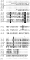
Fig. 1
Fig. 1
Alignment of Anopheles and Drosophila peroxiredoxins. Three of the four major Prx clades are represented: typical 2-Cys and 1-Cys and atypical 2-Cys (PrxV). Not shown are the BCP proteins because they were not identified in these insect genomes. Using the ClustalW method in conjunction with data from [29], the sequences were aligned and residues conserved among all three Prx types were shaded in black. Residues conserved in each clade are in bold. Redox-active cysteines are indicated with a black arrow (▼); residues proposed to be involved in the activation of the peroxidatic cysteine are identified with an open circle (○); residues believed to be involved in maintaining the dimeric structure of Prx are marked with a closed circle (●) [31]; residues highlighted in gray make up the signature sequence for 2-Cys Prx that is sensitive to hyperoxidation [30]. Abbreviations: Ag, Anopheles gambiae; As, Anopheles stephensi; and Dm, Drosophila melanogaster. The prefix “p” indicates that only a partial sequence was obtained.
Fig. 2
Fig. 2
Southern blot analysis of the AsPrx-4783 gene. Genomic DNA was isolated from adult A. stephensi and 12 μg of purified genomic DNA was digested with EcoRI (lane 1), KpnI (lane 2), XhoI (lane 3), or XmnI (lane 4); electrophoretically separated through 0.8% agarose; and transferred onto nylon membrane. The filter was hybridized with the 32P-labeled AsPrx-4783 coding region probe as described under Materials and methods. Molecular size markers (bp) are indicated on the left.
Fig. 3
Fig. 3
(A) Anti-DmPrx-4783 antiserum recognizes DmPrx-4783 and AsPrx-4783. Crude protein lysates (10 μg) from D. melanogaster S2 cells or from A. stephensi MSQ43 cells were subjected to 12% SDS–PAGE under reducing conditions (DTT), transferred to Immobilon-P membrane, and incubated with rabbit anti-DmPrx-4783 antiserum. Treatment conditions: (lane 1) untreated S2 cells, (lane 2) S2 cells stimulated with copper(II) sulfate for 24 h, (lane 3) untreated MSQ43 cells, (lane 4) MSQ43 cells mock transfected for 48 h, (lane 5) MSQ43 cells transfected with TLP58 for 48 h, and (lane 6) MSQ43 cells transfected with AsPrx-4783 dsRNA for 48 h. Molecular weight markers (kDa) are shown on the left. (B) Anti-V5 detects AsPrx-4783 overexpression and dimerization in D. melanogaster S2 cells. S2 cells were collected at various times after mock transfection or transfection with pTLP55, which encodes AsPrx-4783. Crude protein lysates (20 μg per lane) were electrophoretically separated through a 12% SDS–PAGE gel under reducing (with DTT) or nonreducing (without DTT) conditions, transferred to membrane, and probed with anti-V5 antiserum. Proteins with the expected masses of monomer and dimer are indicated. Anti-V5 antibody did not cross-react with proteins in cell extracts prepared from mock-transfected cells. Molecular weight markers (kDa) are shown on the left. (C) Anti-DmPrx-4783 detects AsPrx-4783 overexpression and mixed dimer formation in D. melanogaster S2 cells. Protein samples were isolated at 24 h after copper(II) sulfate treatment from S2 cells that were mock-transfected or transfected with plasmid TLP55. Crude protein lysates (20 μg per lane) were electrophoretically separated through a 12% SDS–PAGE gel under reducing (with DTT) or nonreducing (without DTT) conditions, transferred to membrane, and incubated with anti-DmPrx-4783 antiserum. Cross-reacting proteins correspond to possible AsPrx-4783 dimer and mixed AsPrx-4783/DmPrx-4783 dimer (a), endogenous dimeric DmPrx-4783 (b), fast migrating DmPrx-4783 (c), recombinant monomeric AsPrx-4783 (d), and endogenous monomeric DmPrx-4783 (e). Molecular weight markers (kDa) are shown on the left. (D) Anti-V5 detects AsPrx-4783 overexpression and dimerization in A. stephensi MSQ43 cells. MSQ43 cells were collected at various times after mock transfection or transfection with plasmid TLP58 encoding AsPrx-4783. Crude protein lysates (20 μg per lane) were electrophoretically separated through a 10% SDS–PAGE gel under nonreducing conditions, transferred to membrane, and probed with anti-V5 antibody. Proteins with the expected masses of monomer and dimer are indicated. Molecular weight markers (kDa) are shown on the left.
Fig. 4
Fig. 4
Overexpressed AsPrx-4783 protects transfected D. melanogaster S2 cells from RNOS-specific cytotoxicity. D. melanogaster S2 cells were mock transfected (control cells) or transfected with the pTLP55 plasmid encoding AsPrx-4783 (PRX cells). Protection due to overexpression of AsPrx-4783 was assessed by trypan blue exclusion assay for cell viability. Cells were challenged with (A) 0, 10, or 20 mM hydrogen peroxide, (B) 0, 1500, or 3000 μM DEA-NO, (C) 0, 200, or 500 μM peroxynitrite, and (D) 0, 1, or 2 mM Angeli's salt as described under Materials and methods. Values represent means ± SEM of three independent replicates. Significance versus control is designated: *p < 0.05; Student's t test.
Fig. 5
Fig. 5
Overexpressed AsPrx-4783 protects transfected A. stephensi MSQ43 cells from RNOS-specific cytotoxicity. A. stephensi MSQ43 cells were mock transfected without plasmid (control cells) or transfected with the pTLP58 plasmid encoding AsPrx-4783 (PRX cells). Protection due to overexpression of AsPrx-4783 was assessed by trypan blue exclusion assay for cell viability. Cells were challenged with (A) 0, 125, or 250 μM hydrogen peroxide, (B) 0, 300, or 700 μM DEA-NO, (C) 0, 75, or 150 μM peroxynitrite, and (D) 0, 50, or 100 μM Angeli's salt as described under Materials and methods. Values are the means ± SEM of the results from three independent replicates. Significance versus control is designated: *p < 0.05; Student's t test.
Fig. 6
Fig. 6
dsRNA-mediated AsPrx-4783 silencing in A. stephensi MSQ43 cells is specific and persistent. MSQ43 cell cultures were collected 24–120 h after mock transfection (no dsRNA) or transfection with double-stranded RNA for AsPrx-4783 (dsPRX) or murine cyclophilin (dsCyc). Total RNA was isolated (TRIzol) and cDNA was synthesized using oligo(dT)16 (Applied Biosystems) for use in two-step RT-PCR. Relative AsPrx-4783 transcript levels were normalized to AsS7. Relative levels of AsPrx-4783 are shown as normalized ratios in cells transfected with dsPrx or dsCyc; no impact of transfection on AsPrx-4783 would be equivalent to a relative expression level of 1.0. Addition of 400 ng dsPRX resulted in an 81–96% decrease in AsPrx-4783 transcript levels relative to both dsCyc and no dsRNA controls.
Fig. 7
Fig. 7
Silencing of AsPrx-4783 increased susceptibility of A. stephensi MSQ43 cells to RNOS-specific cytotoxicity. A. stephensi MSQ43 cells were mock transfected (control) or transfected with double-stranded AsPrx-4783 RNA (dsPRX). After transfection, cells were challenged with (A) 0, 125, or 250 μM hydrogen peroxide, (B) 0, 300, or 700 μM DEA-NO, (C) 0, 75, or 150 μM peroxynitrite, and (D) 0, 50, or 100 μM Angeli's salt as described under Materials and methods. Cell viability was assessed by the trypan blue exclusion assay. Values represent means ± SEM from three independent replicates. Significance versus control is designated: *p < 0.05; Student's t test.
Fig. 8
Fig. 8
Northern blotting analysis and qRT-PCR of AsPrx-4783 expression in vivo. (A) Messenger RNA was collected from whole non-blood-fed (NB), P. berghei-infected (I), and uninfected (U) A. stephensi at 1 h and 7 days after blood feeding. Aliquots of 9 μg mRNA were separated on a 1% agarose NorthernMax (Ambion) gel and transferred to a nylon membrane. RNA probe was prepared from cloned template containing full-length coding sequence for AsPrx-4783. The cross-hybridizing AsPrx-4783 transcript has an estimated size of 1194 bp, which corresponds to the predicted transcript size of 1223 bp based on sequence analysis. Densitometry was used to quantitate transcript abundance. AsPrx-4783 transcript levels were normalized against AsS7 ribosomal protein gene transcript levels to correct for loading differences; these values are indicated at the bottom. (B, C) Quantitative RT-PCR as described under Materials and methods was performed on total RNA prepared from midguts, carcasses (body tissues minus midguts), and whole bodies of non-blood-fed (NB) mosquitoes and from the same tissues of mosquitoes after ingestion of an uninfected (UN) or P. berghei-infected (INF) blood meal. A single cohort of mosquitoes was used to determine the effects of blood feeding alone (B), whereas four cohorts of mosquitoes with overlapping time points were used to determine the effects of parasite infection on AsPrx-4783 expression (C). Relative induction of AsPrx-4783 expression in the A. stephensi midgut is shown as the ratio of UN to NB expression (B) and as the ratio of INF to UN expression (C). No induction of AsPrx-4783 expression was apparent in the carcass or whole body after blood feeding or infection (not shown).

Similar articles

Cited by

References

    1. Fang FC, editor. Nitric oxide and infection. Kluwer Academic/Plenum; New York: 1999.
    1. Bryk R, Griffin P, Nathan C. Peroxynitrite reductase activity of bacterial peroxiredoxins. Nature. 2000;407:211–215. - PubMed
    1. Chen L, Xie QW, Nathan C. Alkyl hydroperoxide reductase subunit C (AhpC) protects bacterial and human cells against reactive nitrogen intermediates. Mol Cell. 1998;1:795–805. - PubMed
    1. Wong CM, Zhou Y, Ng RW, Kung HF, Jin DY. Cooperation of yeast peroxiredoxins Tsa1p and Tsa2p in the cellular defense against oxidative and nitrosative stress. J Biol Chem. 2002;277:5385–5394. - PubMed
    1. Dubuisson M, Vander Stricht D, Clippe A, Etienne F, Nauser T, Kissner R, Koppenol WH, Rees JF, Knoops B. Human peroxiredoxin 5 is a peroxynitrite reductase. FEBS Lett. 2004;571:161–165. - PubMed

Publication types

MeSH terms

LinkOut - more resources