. 2006 Mar 15;26(11):2862-70.
doi: 10.1523/JNEUROSCI.4048-05.2006.

Glial cells dilate and constrict blood vessels: a mechanism of neurovascular coupling

Affiliations

Glial cells dilate and constrict blood vessels: a mechanism of neurovascular coupling

Monica R Metea et al. J Neurosci. .

Abstract

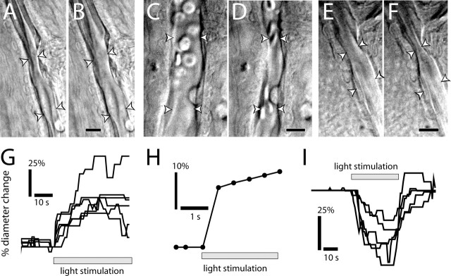
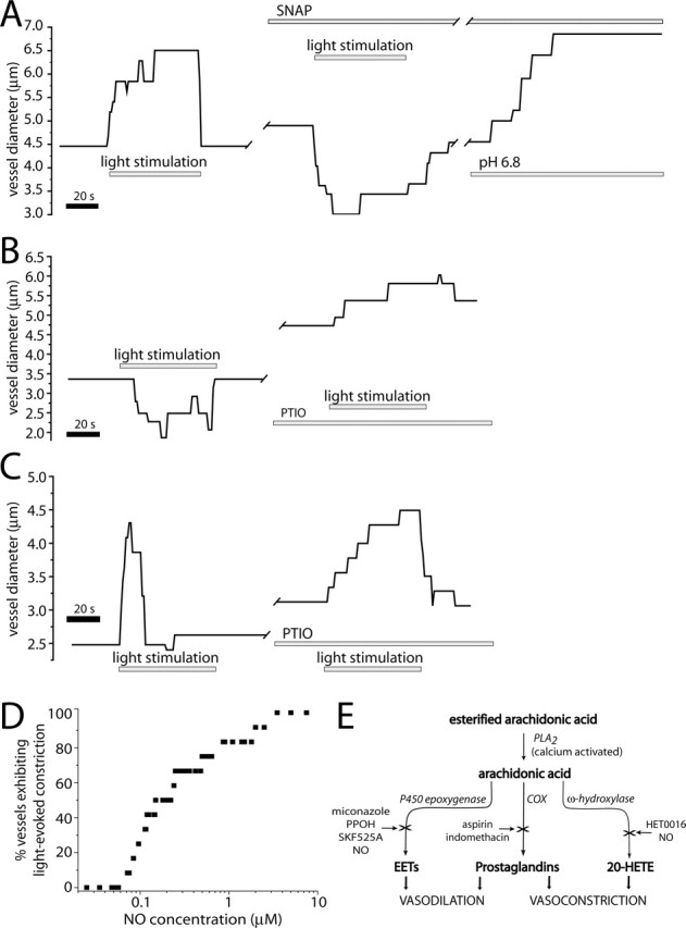
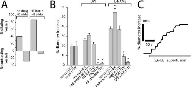
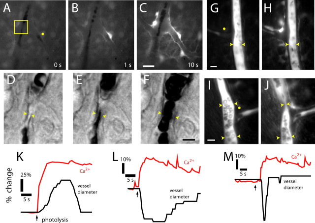
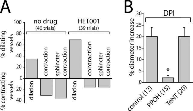
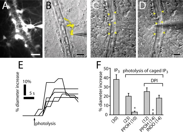
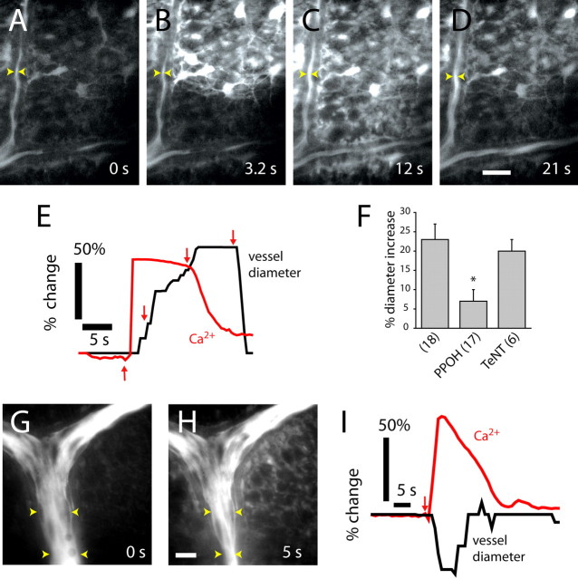
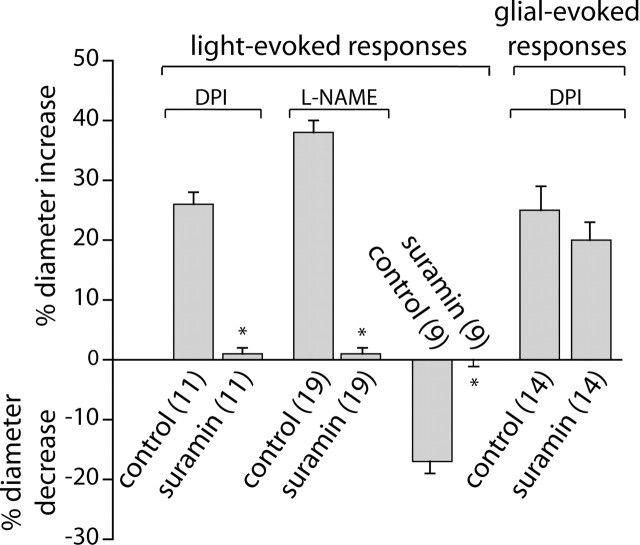
Neuronal activity evokes localized changes in blood flow. Although this response, termed neurovascular coupling, is widely used to monitor human brain function and diagnose pathology, the cellular mechanisms that mediate the response remain unclear. We investigated the contribution of glial cells to neurovascular coupling in the acutely isolated mammalian retina. We found that light stimulation and glial cell stimulation can both evoke dilation or constriction of arterioles. Light-evoked and glial-evoked vasodilations were blocked by inhibitors of cytochrome P450 epoxygenase, the synthetic enzyme for epoxyeicosatrienoic acids. Vasoconstrictions, in contrast, were blocked by an inhibitor of omega-hydroxylase, which synthesizes 20-hydroxyeicosatetraenoic acid. Nitric oxide influenced whether vasodilations or vasoconstrictions were produced in response to light and glial stimulation. Light-evoked vasoactivity was blocked when neuron-to-glia signaling was interrupted by a purinergic antagonist. These results indicate that glial cells contribute to neurovascular coupling and suggest that regulation of blood flow may involve both vasodilating and vasoconstricting components.


Figures

Figure 1.
Figure 1.
Light evokes vasodilation and vasoconstriction. A–F, IR-DIC images of arterioles at the vitreal surface of the retina. Shown are vessels before (A) and during (B) light-evoked vasodilation, before (C) and during (D) light-evoked constriction, and before (E) and during (F) light-evoked sphincter-like constriction. Scale bars, 10 μm. Arrowheads in all figures indicate the diameter of the vessel lumen. G, Time course of light-evoked vasodilation in six trials. H, Time course of vasodilation in a rapidly responding vessel (latency, <500 ms). I, Time course of vasoconstriction in five trials.
Figure 2.
Figure 2.
NO modulates light-evoked vasomotor responses. A, Light-evoked vasodilation is transformed into a vasoconstriction by addition of the NO donor SNAP (100 μm). The vessel dilates in the presence of SNAP and responds to a decrease in pH of the superfusate with an additional dilation. B, Light-evoked vasoconstriction is transformed into a vasodilation by addition of the NO scavenger PTIO (100 μm). The vessel dilates in the presence of PTIO. C, PTIO increases the duration of a light-evoked vasodilation. D, Raising NO levels increases the percentage of vessels that constrict to light stimulation. At <70 nm NO, light stimulation evoked vasodilation in all vessels (n = 12). As NO was raised, a greater percentage of vessels constricted. E, Schematic of arachidonic acid metabolism, showing synthesis of vasoactive EETs, prostaglandins, and 20-HETE. Synthetic enzymes are italicized and enzyme inhibition is indicated by an X.
Figure 3.
Figure 3.
Arachidonic acid metabolites mediate light-evoked vasomotor responses. A, The ω-hydroxylase inhibitor HET0016 (100 nm) decreases the percentage of light-evoked vasoconstrictions and increases the percentage of vasodilations. B, Pharmacology of light-evoked vasodilation. In the presence of the NOS inhibitors DPI (3 μm) or L-NAME (100 μm), the epoxygenase inhibitors PPOH (20 μm), SKF525A (20 μm), and miconazole (10 μm) reduce light-evoked vasodilation. The COX inhibitors aspirin (50 μm) and indomethacin (5 μm), in contrast, do not reduce vasodilation. In all figures, asterisks indicate a significant difference from controls (p < 0.05; Student’s t test). Error bars indicate SEM. The number of samples is given in parentheses. C, Superfusion of 5,6-EET (1 μm) increases the diameter of an arteriole.
Figure 4.
Figure 4.
Photolysis of caged Ca2+ in glial cells evokes vasodilation and vasoconstriction. A–C, Fluorescence images showing a Ca2+ increase that propagates through several glial cells after an uncaging flash. D–F, Glial-evoked vasodilation. IR-DIC images of the region indicated in A are shown, each acquired 0.5 s after the corresponding image above. G, H, Glial-evoked vasoconstriction. Fluorescence images of a fluorescein-filled vessel before and after photolysis. Note the photolysis-evoked increase in glial Ca2+. I, J, Glial-evoked sphincter-like vasoconstriction. Fluorescence images of a fluorescein-filled vessel before and after photolysis. Yellow dots in A, G, and I indicate sites of uncaging flash. Scale bars: C, 20 μm; F, 5 μm; G, 10 μm; I, 10 μm. K–M, Time course of glial Ca2+ change and vessel dilation (K), constriction (L), and sphincter-like constriction (M).
Figure 5.
Figure 5.
Arachidonic acid metabolites mediate glial-evoked vasomotor responses. A, The ω-hydroxylase inhibitor HET0016 (100 nm) decreases the percentage of photolysis-evoked vasoconstrictions and increases the percentage of vasodilations. B, In the presence of DPI (3 μm), photolysis of caged Ca2+ evokes vasodilation that is blocked by the epoxygenase inhibitor PPOH (20 μm) but not by preincubation with TeNT.
Figure 6.
Figure 6.
IP3-stimulated glial cells evoke vasodilation. A, Lucifer yellow-filled astrocytes are shown in a fluorescence image. Multiple astrocyte processes envelop an arteriole. B, A Lucifer yellow-filled Müller cell contacting an arteriole is shown in a superimposed fluorescence and IR-DIC image. C, D, Arteriole dilation evoked by introduction of IP3 into a Müller cell. A vessel is shown before (C) and during (D) whole-cell recording with a pipette containing IP3 (250 μm). Scale bars: A, B, D, 20 μm. E, Photolysis of caged IP3 (250 μm) in Müller cells and astrocytes evokes vasodilation. F, Introduction of IP3 into glial cells evokes vasodilation. Photolysis of caged IP3 evokes vasodilation that is reduced by PPOH (20 μm) but not indomethacin (5 μm).
Figure 7.
Figure 7.
Propagated glial Ca2+ waves evoke vasodilation and vasoconstriction in distant arterioles. A–D, Fluorescence images showing an ATP-evoked Ca2+ wave propagating through glial cells at the vitreal surface of the retina. Dilation occurs when the Ca2+ wave reaches the vessel. The site of focal ATP ejection, used to initiate the Ca2+ wave, was just beyond the top right corner of the images. Scale bar, 20 μm. E, Time course of Ca2+ increase and vessel dilation in A–D, measured at the arrowheads in the images. Arrows in E indicate the time of acquisition of images A–D. F, Vessel dilation after arrival of glial Ca2+ waves is reduced by the epoxygenase inhibitor PPOH (20 μm) but not by preincubation in TeNT. L-NAME (100 μm) or DPI (3 μm) is present in superfusate. G, H, Fluorescence images before and after the arrival of a glial Ca2+ wave. Vasoconstriction occurs after the arrival of the Ca2+ wave. The vessel is filled with fluorescein. Scale bar, 20 μm. I, Time course of Ca2+ increase and vessel constriction after initiation (arrow) of a Ca2+ wave.
Figure 8.
Figure 8.
Suramin blocks light-evoked but not glial-evoked vasomotor responses. In the presence of DPI (3 μm) or L-NAME (100 μm), light-evoked vasodilations are almost completely blocked by 100 μm suramin. Light-evoked vasoconstrictions are completely abolished by suramin. In contrast, glial-evoked vasodilations are not reduced by suramin. Glial cells were stimulated by photolysis of caged IP3.

References

    1. Alkayed NJ, Narayanan J, Gebremedhin D, Medhora M, Roman RJ, Harder DR (1996). Molecular characterization of an arachidonic acid epoxygenase in rat brain astrocytes. Stroke 27:971–979. - PubMed
    1. Alonso-Galicia M, Sun CW, Falck JR, Harder DR, Roman RJ (1998). Contribution of 20-HETE to the vasodilator actions of nitric oxide in renal arteries. Am J Physiol Renal Physiol 275:F370–F378. - PubMed
    1. Alonso-Galicia M, Falck JR, Reddy KM, Roman RJ (1999). 20-HETE agonists and antagonists in the renal circulation. Am J Physiol Renal Physiol 277:F790–F796. - PubMed
    1. Amruthesh SC, Boerschel MF, McKinney JS, Willoughby KA, Ellis EF (1993). Metabolism of arachidonic acid to epoxyeicosatrienoic acids, hydroxyeicosatetraenoic acids, and prostaglandins in cultured rat hippocampal astrocytes. J Neurochem 61:150–159. - PubMed
    1. Anderson CM, Nedergaard M (2003). Astrocyte-mediated control of cerebral microcirculation. Trends Neurosci 26:340–344. - PubMed

Publication types

MeSH terms

Substances

LinkOut - more resources