. 2006 Mar 15;26(11):2923-32.
doi: 10.1523/JNEUROSCI.4390-05.2006.

Genetic manipulation of intraspinal plasticity after spinal cord injury alters the severity of autonomic dysreflexia

Affiliations

Genetic manipulation of intraspinal plasticity after spinal cord injury alters the severity of autonomic dysreflexia

Adrian A Cameron et al. J Neurosci. .

Abstract

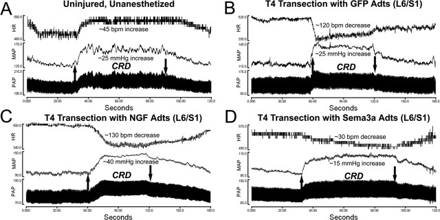
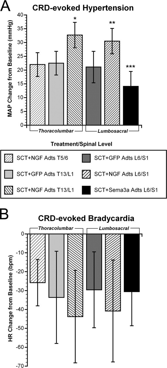
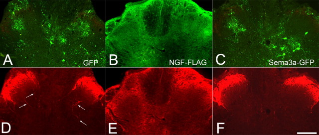
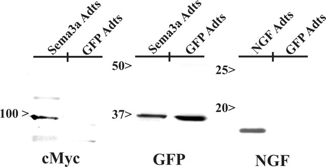
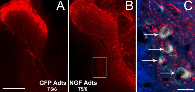
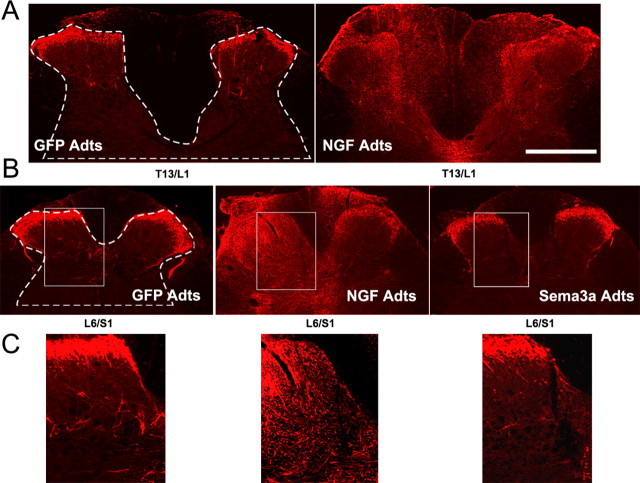
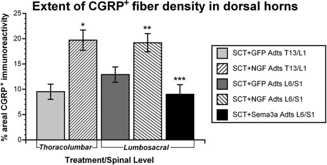
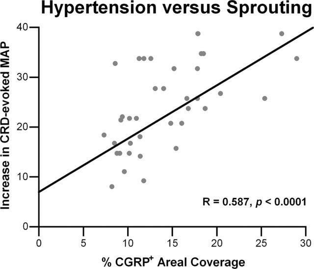
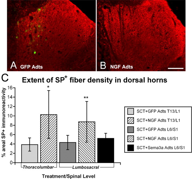
Severe spinal cord injuries above mid-thoracic levels can lead to a potentially life-threatening hypertensive condition termed autonomic dysreflexia, which is often triggered by painful distension of pelvic viscera (bladder or bowel) and consequent sensory fiber activation, including nociceptive C-fibers. Interruption of tonically active medullo-spinal pathways after injury causes disinhibition of thoracolumbar sympathetic preganglionic neurons, and intraspinal sprouting of nerve growth factor (NGF)-responsive primary afferent fibers is thought to contribute to their hyperactivity. We investigated spinal levels that are critical for eliciting autonomic dysreflexia using a model of noxious colorectal distension (CRD) after complete spinal transection at the fourth thoracic segment in rats. Post-traumatic sprouting of calcitonin gene-related peptide (CGRP)-immunoreactive primary afferent fibers was selectively altered at specific spinal levels caudal to the injury with bilateral microinjections of adenovirus encoding the growth-promoting NGF or growth-inhibitory semaphorin 3A (Sema3a) compared with control green fluorescent protein (GFP). Two weeks later, cardio-physiological responses to CRD were assessed among treatment groups before histological analysis of afferent fiber density at the injection sites. Dysreflexic hypertension was significantly higher with NGF overexpression in lumbosacral segments compared with GFP, whereas similar overexpression of Sema3a significantly reduced noxious CRD-evoked hypertension. Quantitative analysis of CGRP immunostaining in the spinal dorsal horns showed a significant correlation between the extent of fiber sprouting into the spinal segments injected and the severity of autonomic dysreflexia. These results demonstrate that site-directed genetic manipulation of axon guidance molecules after complete spinal cord injury can alter endogenous circuitry to modulate plasticity-induced autonomic pathophysiology.


Figures

Figure 1.
Figure 1.
A–D, Illustrative traces of pulsatile arterial pressure (PAP), MAP, and HR before, during, and after 1 min of noxious CRD in uninjured awake animals (A) versus injured rats 15 d after injury with bilateral injections into L6/S1 with adenovirus encoding GFP (B), NGF (C), or Sema3a (D). Upward and downward arrows indicate rectal balloon catheter inflation and deflation, respectively. Note that in uninjured rats, the HR and MAP rise and fall in synchrony in response to CRD, whereas all injured animals show autonomic dysreflexia with hypertension accompanied by bradycardia.
Figure 2.
Figure 2.
Quantitative assessments of autonomic dysreflexia severity in injured rats among treatment groups. A, Two weeks after T4 spinal cord transection (SCT), NGF overexpression in either T13/L1 or L6/S1 significantly increased hypertension versus GFP overexpression at the same levels, as well as NGF Adts injections at T5/6. Conversely, Sema3a overexpression in L6/S1 significantly reduced noxious CRD-induced hypertension. B, The bradycardia manifested in response to visceral pain in all rats with SCT was significantly (p < 0.0001) different from and diametrically opposite to tachycardia observed in uninjured rats (data not shown). The magnitude of bradycardia across the groups mirrored the extent of hypertension, despite the high variability in HR within treatment groups. Bars represent means ± SD. *p < 0.01 versus GFP Adts T13/L1 and NGF Adts T5/6; **p < 0.005 versus GFP Adts L6/S1; ***p < 0.01 versus GFP Adts L6/S1 and p < 0.0001 versus NGF Adts L6/S1.
Figure 3.
Figure 3.
A–C, Series of photomicrographs demonstrating how sections were designated for CGRP+ fiber immunostaining and quantification at sites of bilateral adenovirus microinjections that encoded control GFP (A), NGF tagged with the FLAG epitope (B), or Sema3a that coexpressed GFP (C). The GFP localization was intracellular, particularly in glial and neuronal subpopulations, whereas exogenous FLAG immunoreactivity appeared as widespread extracellular staining. Dual immunofluorescent images in the bottom row show the corresponding spatial extent of CGRP+ immunostaining in L6/S1 segments with overexpression of GFP (D), NGF (E), or Sema3a (F). Note the modest CGRP+ fiber sprouting seen in GFP controls (arrows in D). Conversely, the widespread and punctate immunostaining of CGRP+ fibers mirrored diffuse immunolocalization of exogenous NGF–FLAG throughout the dorsal horns and even dorsal columns. Scale bar: (in F) A–F, 200 μm.
Figure 4.
Figure 4.
Western blot analyses confirm increased expression of transgene protein after injections of Sema3a Adts, GFP Adts, or NGF Adts into spinal cords. Sema3a was identified at ∼97 kDa using an anti-myc antibody to recognize this C-terminal tag on the protein. GFP expression was observed in both Sema3a Adts- and control-injected cords. Likewise, NGF (17 kDa) was identified using a antibody specific for NGF.
Figure 5.
Figure 5.
Photomicrographs showing CGRP+ fiber immunostaining at mid-thoracic spinal levels that received bilateral Adts injections. A, B, Compared with control GFP Adts injections (A), the overexpression of NGF at T5/6 (B) elicited profuse CGRP+ fiber sprouting into the dorsal spinal cord after 15 d postinjury. C, Superimposed dual-immunofluorescence, high-magnification images of NGF Adts-injected lateral gray matter (boxed region in B) shows numerous CGRP+ fibers extending proximal to sympathetic preganglionic neurons (arrows) prelabeled with FluoroGold. This was not observed in T5/6 cords injected with GFP Adts (data not shown). Scale bars: (in A, C) A, B, 200 μm; C, 20 μm.
Figure 6.
Figure 6.
A, B, Photomicrographs showing CGRP+ fiber immunostaining at thoracolumbar (A) or lumbosacral (B) spinal levels that received bilateral Adt injections. C, Higher magnifications demonstrate CGRP+ immunostaining in L6/S1 dorsal horns (boxed regions in B). Compared with control GFP Adts, NGF overexpression in the thoracolumbar and lumbosacral spinal cord induced CGRP+ fiber sprouting throughout the dorsal horns and even dorsal columns. Conversely, Sema3a Adts reduced post-traumatic aberrant CGRP+ fiber sprouting into the L6/S1 dorsal horns. Scale bar: (in A) A, B, 500 μm; C, 200 μm.
Figure 7.
Figure 7.
Quantitative spatial analysis of CGRP+ fiber staining in dorsal horns (see dashed lines in Fig. 6 A,B) of sections through Adts injection sites 15 d after spinal cord transection (SCT). NGF overexpression in thoracolumbar and lumbosacral regions significantly increased CGRP+ fiber density compared with control GFP. In contrast, Sema3a Adts overexpression in lumbosacral levels significantly reduced CGRP+ fiber density versus injured controls. Bars represent means ± SD. *p < 0.001 versus GFP Adts T13/L1; **p < 0.005 versus GFP Adts L6/S1; ***p < 0.001 versus GFP Adts L6/S1 and p < 0.0001 versus NGF Adts L6/S1.
Figure 8.
Figure 8.
Linear regression analysis demonstrates a significant (p < 0.0001) positive correlation between the extent of noxious CRD-evoked hypertension and the density of CGRP+ fibers in dorsal horns among spinal rats overexpressing GFP, NGF, or Sema3a at the different spinal levels.
Figure 9.
Figure 9.
A, B, Photomicrographs showing SP+ fiber staining in thoracolumbar dorsal horns injected with adenovirus encoding control GFP Adts (A) versus NGF Adts (B). Note transfected cells expressing GFP seen with dual immunofluorescence in A. Similar to CGRP+ fiber staining, NGF overexpression elicited profuse SP+ fiber sprouting throughout the dorsal horns, although the staining is far less intense and more punctuate in this CGRP+ fiber subpopulation. Scale bar: (in B) 200 μm. C, Quantitative spatial analysis of SP+ immunostaining in dorsal horns of sections adjacent to those stained for CGRP 15 d after spinal cord transection (SCT). NGF overexpression significantly increased SP+ fiber density compared with control GFP. In contrast, with Sema3a overexpression, there was no observable alteration in SP+ fiber densities versus injured controls. Bars represent means ± SD. *p < 0.01 versus GFP Adts T13/L1; **p < 0.05 versus GFP Adts L6/S1 and Sema3a Adts L6/S1.

References

    1. Anderson CR, Edwards SL (1994). Intraperitoneal injections of Fluorogold reliably labels all sympathetic preganglionic neurons in the rat. J Neurosci Methods 53:137–141. - PubMed
    1. Anderson KD (2004). Targeting recovery: priorities of the spinal cord injured population. J Neurotrauma 21:1371–1383. - PubMed
    1. Averill S, McMahon SB, Clary DO, Reichardt LF, Priestley JV (1995). Immunocytochemical localization of trkA receptors in chemically identified subgroups of adult rat sensory neurons. Eur J Neurosci 7:1484–1494. - PMC - PubMed
    1. Bennett AD, Taglialatela G, Perez-Polo R, Hulsebosch CE (1999). NGF levels decrease in the spinal cord and dorsal root ganglion after spinal hemisection. NeuroReport 10:889–893. - PubMed
    1. Bennett DL, Dmietrieva N, Priestley JV, Clary D, McMahon SB (1996). trkA, CGRP and IB4 expression in retrogradely labelled cutaneous and visceral primary sensory neurones in the rat. Neurosci Lett 206:33–36. - PubMed

Publication types

MeSH terms