Comparative Study
. 2006 Mar 15;26(11):2971-80.
doi: 10.1523/JNEUROSCI.5453-05.2006.

Regulation of dendritic branching and spine maturation by semaphorin3A-Fyn signaling

Affiliations
Comparative Study

Regulation of dendritic branching and spine maturation by semaphorin3A-Fyn signaling

Asa Morita et al. J Neurosci. .

Abstract

A member of semaphorin family, semaphorin3A (Sema3A), acts as a chemorepellent or chemoattractant on a wide variety of axons and dendrites in the development of the nervous systems. We here show that Sema3A induces clustering of both postsynaptic density-95 (PSD-95) and presynaptic synapsin I in cultured cortical neurons without changing the density of spines or filopodia. Neuropilin-1 (NRP-1), a receptor for Sema3A, is present on both axons and dendrites. When the cultured neurons are exposed to Sema3A, the cluster size of PSD-95 is markedly enhanced, and an extensive colocalization of PSD-95 and NRP-1 or actin-rich protrusion is seen. The effects of Sema3A on spine morphology are blocked by PP2, an Src type tyrosine kinase inhibitor, but not by the PP3, the inactive-related compound. In the cultured cortical neurons from fyn(-/-) mice, dendrites bear few spines, and Sema3A does not induce PSD-95 cluster formation on the dendrites. Sema3A and its receptor genes are highly expressed during the synaptogenic period of postnatal days 10 and 15. The cortical neurons in layer V, but not layer III, show a lowered density of synaptic bouton-like structure on dendrites in sema3A- and fyn-deficient mice. The neurons of the double-heterozygous mice show the lowered spine density, whereas those of single heterozygous mice show similar levels of the spine density as the wild type. These findings suggest that the Sema3A signaling pathway plays an important role in the regulation of dendritic spine maturation in the cerebral cortex neurons.


Figures
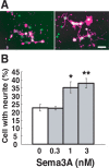
Figure 1.
Figure 1.
Sema3A induces neurite formation in cortical neurons. A, Cultured E16.5 mouse cortical neurons on 1 DIV were applied with Sema3A (left, 0 nm; right, 1 nm) and further incubated for 24 h. Neurons were double stained with rhodamine-labeled phalloidin (purple) and DAPI (green). Scale bar, 50 μm. B, Neurite formation was quantified by scoring the number of cells possessing neurites and expressing them as a percentage of the totals. Neurites were defined as process extensions greater than one cell body in length. Each value represents mean ± SEM of 20 neurons from four independent cultures. *p < 0.005, ** p < 0.001 compared with vehicle control. The statistical significance of the results was analyzed using one-way ANOVA.
Figure 2.
Figure 2.
Sema3A induces dendritic branching in cortical neurons. A, Morphological changes of the cultured neurons from E16.5 mouse on 7 DIV. The wild-type cortical neurons applied with (b, d) and without (a, c) Sema3A (3 nm) and the fyn-deficient cortical neurons applied with (f) and without (e) Sema3A (3 nm) were immunostained by anti-MAP2 antibody. Lavendustin A (3 μm), a tyrosine kinase inhibitor (c, d), or vehicle control (a, b) was applied to the cultured neurons 30 min before application of Sema3A. Scale bar, 20 μm. B, Quantitative analysis of Sema3A-induced branching. Total numbers of branch points were estimated for 20 neurons from four independent cultures. C, Quantitative analysis of suppressive effect of lavendustin A (3 μm), PP2 (0.4 μm), or PP3 (0.4 μm) on the Sema3A-induced branching. **p < 0.01 compared with corresponding control. D, Quantitative analysis of Sema3A-induced branching in fyn-deficient mouse neurons. wt, Wild type. *p < 0.05, **p < 0.001 compared with vehicle control or Sema3A (3 nm) alone. Statistical significance was assessed with a one-way ANOVA.
Figure 3.
Figure 3.
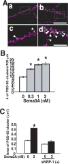
Sema3A increases PSD-95 immunoreactive clusters in cultured cortical neurons. A, The cultured E16.5 cortical neurons on 14 DIV were applied with (b, d) or without (a, c) Sema3A (3 nm) and were double stained with anti-PSD-95 (green) and rhodamine–phalloidin (purple). Magnified images of boxed area in a and b are shown in c and d, respectively. White color indicates localization of PSD-95 clusters on actin-rich protrusions stained with rhodamine–phalloidin on dendrites (arrowheads). Scale bars: b, 10 μm; d, 5 μm. B, Sema3A concentration dependently promoted the density of PSD-95-immunoreactive clusters per 100 μm of dendritic shaft, estimated from 20 neurons from four independent cultures. *p < 0.001 compared with vehicle control. C, Quantitative analysis of area size of PSD-95 clusters in culture treated with or without Sema3A. sNRP-1 (30 nm) suppressed the promoting effect of Sema3A on PSD-95 clusters (n = 78–87 from two independent cultures). *p < 0.001 compared with vehicle control. The immunofluorescence was detected by confocal microscopy, and the areas were quantified by LSM-5 image software (Zeiss).
Figure 4.
Figure 4.
Colocalization of clusters of PSD-95 and NRP-1 on spine-like protrusions on dendrites. The cultured cortical neurons were applied with (b, d) or without (a, c) Sema3A (3 nm) and were double stained with anti-PSD-95 (purple) and NRP-1 (green) antibodies. Magnified images of boxed area in a and b are shown in c and d, respectively. The white color indicates colocalization of PSD-95 and NRP-1 clusters (arrowheads). Scale bars: a, b, 10 μm; c, d, 5 μm.
Figure 5.
Figure 5.
Src-type tyrosine kinase inhibitors block Sema3A-induced PSD-95 clustering. PP2 but not PP3 suppressed the effect of Sema3A (3 nm) on PSD-95 clusters, and Sema3A fails to induce PSD-95 cluster formation in fyn−/− neurons. A, PSD-95 clusters in cultured cortical neurons treated with (b, d) and without (a, c) Sema3A. PP2 (a, b) or PP3 (c, d) (0.4 μm) was applied 30 min before Sema3A. Scale bar, 10 μm. B, Quantitative analysis of the effects of PP2 and PP3 on the density of PSD-95 clusters. Images of 10 typical neurons from two independent culture preparations were recorded, and the density of PSD-95-clusters was measured. C, Quantitative analysis of the effects of PP2 and PP3 on the area of PSD-95 clusters. Images of 10 typical neurons from two independent culture preparations were recorded, and the area of PSD-95 clusters was measured. D, Quantitative analysis of the effect of lavendustin A on the density of PSD-95 clusters. E, The cultured cortical neurons from fyn−/− mice were applied with (b) or without (a) Sema3A (3 nm) and were double stained with anti-PSD-95 (green) and rhodamine–phalloidin (purple). Scale bar, 10 μm. F, There were few dendritic spine-like protrusions in fyn−/− neurons, and Sema3A induced neither PSD-95 clustering nor spine-like protrusion on dendrites. *p < 0.001 compared with Sema3A at 0 nm; p < 0.001 compared with corresponding value in wild type (wt).
Figure 6.
Figure 6.
Sema3A induces cluster formation of PSD-95 and synapsin I on actin-rich protrusions. A, Sema3A increased synapsin I-immunoreactive clusters in cortical neurons. Cultured E16.5 mouse cortical neurons on 14 DIV were treated with (b, d) or without (a, c) Sema3A (3 nm) for 24 h. Cortical neurons were stained with anti-synapsin I antibody. Magnified images of boxed area in a and b are shown in c and d, respectively. Scale bars, 10 μm. B, Sema3A increased the density of synapsin I clusters (green) in apposition of the PSD-95 clusters (purple) in the cortical neurons. The cultured cortical neurons were treated with (b) or without (a) Sema3A (3 nm) for 24 h. The neurons were double stained with anti-synapsin I and anti-PSD95 antibodies. Superimposition of images, illustrating clusters of PSD-95-immunoreactive spots, were closely associated with those of synapsin I (arrows). Scale bar, 10 μm. C, Superposition of immunopositive clusters of synapsin I (green) and rhodamine–phalloidin (purple) in cultured neurons with (b) or without (a) Sema3A. Arrowheads indicate synapsin I clusters contacted with rhodamine–phalloidin fluorescent signals. Scale bar, 10 μm.
Figure 7.
Figure 7.
Embryonic and early postnatal expression of nrp-1 and sema3A genes in the cortex. A, Mouse P0 (a), P10 (b), P15 (c), and 4-week-old (d) sagittal sections processed for in situ hybridization with probe for Sema3A and NRP-1. Cortical layers are shown on the left. Scale bar, 100 μm. MZ, Marginal zone; CP, cortical plate; tr, nerve fiber tract; VZ, ventricular zone. B, Immunoblot analysis of P0, P10, P15, and 4-week-old brain lysates with anti-Sema3A antibody and anti-NRP-1 antibodies. In general, Sema3A and NRP-1 were detectable in NP-40-soluble lysates of P0 to 4-week-old brains. To detect Sema3A in the brain at the developmental stage of P0, the pellets obtained were dissolved in 2× the sample buffer and further sonicated (Insoluble).
Figure 8.
Figure 8.
Dendritic spine morphology in cortical neurons in sema3A and fyn−/− mice. A, Photographs of Golgi impregnation of wild-type (a), sema3A−/− (b), and fyn−/− (c) mice. Scale bar, 10 μm. Unbranched dendritic protrusions with a head showing a bouton-like structure were defined as dendritic spines (arrows). B, Number of dendritic spines expressed on basal or apical branches of layer V pyramidal neurons. The number of spine per 100 μm of dendritic shaft was calculated. Each value represents mean ± SEM estimated from 20 individual neurons. wt, Wild type. *p < 0.001 compared with the corresponding value in wild type (wt).

Similar articles

Cited by

References

    1. Ahmari SE, Buchanan J, Smith SJ (2000). Assembly of presynaptic active zones from cytoplasmic transport packets. Nat Neurosci 3:445–451. - PubMed
    1. Aizawa H, Wakatsuki S, Ishii A, Moriyama K, Sasaki Y, Ohashi K, Sekine-Aizawa Y, Sehara-Fujisawa A, Mizuno K, Goshima Y, Yahara I (2001). Phosphorylation of cofilin by LIM-kinase is necessary for semaphorin 3A-induced growth cone collapse. Nat Neurosci 4:367–373. - PubMed
    1. Boda B, Alberi S, Nikonenko I, Node-Langlois R, Jourdain P, Moosmayer M, Parisi-Jourdain P, Muller D (2004). The mental retardation protein PAK3 contributes to synapse formation and plasticity in hippocampus. J Neurosci 24:10816–10825. - PMC - PubMed
    1. Campbell DS, Holt CE (2001). Chemotropic responses of retinal growth cones mediated by rapid local protein synthesis and degradation. Neuron 32:1013–1026. - PubMed
    1. Ethell IM, Irie F, Kalo MS, Couchman JR, Pasquale EB, Yamaguchi Y (2001). EphB/syndecan-2 signaling in dendritic spine morphogenesis. Neuron 31:1001–1013. - PubMed

Publication types

MeSH terms