. 2006 Jun;73(6):767-73.
doi: 10.1002/mrd.20466.

Calpain 11 is unique to mouse spermatogenic cells

Affiliations

Calpain 11 is unique to mouse spermatogenic cells

Irit Ben-Aharon et al. Mol Reprod Dev. 2006 Jun.

Abstract

The calpains are a family of calcium-dependent thiol proteases involved in intracellular processing of proteins. They occur as heterodimers containing one of various large subunits and a common small subunit. Some of the large subunits are expressed ubiquitously and others are expressed in a restricted set of tissues. We have cloned the cDNA for mouse calpain 11 and demonstrated that it is expressed specifically in the mouse testis. The mRNA begins to accumulate in the testis between days 14 and 16 after birth, corresponding to the period of pachytene spermatocyte development. The protein is detected by day 18 after birth, during mid to late pachytene spermatocyte development, and is present in the acrosomal region of spermatozoa from the cauda epididymis. The expression of calpain 11 during spermatogenesis and its localization in spermatozoa suggest that it is involved in regulating calcium-dependent signal transduction events during meiosis and sperm functional processes.


Figures
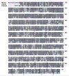
Figure 1
Figure 1
Comparison of the deduced amino acid sequences of mouse CAPN11 with those of human CAPN11 and chicken m/μ calpain. Shaded areas indicate amino acid homology. Brackets define EF-hand motifs.
Figure 2
Figure 2
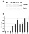
Capn11 mRNA expression. A. Expression of Capn11 mRNA as determined by RT-PCR in mouse brain (B), heart (H), kidney (K) liver (Li), lung (Lu), ovary (O), spleen (S), stomach (St), thymus (T), epididymis (E), mixed germ cells (M), and testis (T). Actin was detected in all tissue samples at approximately equal levels. Images are from an Agilent 2100 Bioanalyzer readout. B. Expression of Capn11 mRNA as determined by real-time RT-PCR in testes from mice at days 10 to 28 after birth. Levels are expressed in terms of fold change compared to day 8.
Figure 3
Figure 3
Developmental expression of CAPN11. A. Testes were collected from 6 to 30 day old mice and crude protein extracts were isolated by homogenizing in lysis buffer. Proteins were resolved by SDS-PAGE analysis and transferred onto ImobilonTM nylon membrane. The blots were incubated with the antiserum (1:100) to the CAPN11 peptide, followed by ECL detection. The 83 kDa CAPN11 band (arrow) is distinct by day 18. B. Preincubating the antiserum with the CAPN11 peptide (1:1) considerably reduced the immunostaining of the 83 kDa CAPN11 band, but not the cross-reaction with the higher molecular weight band.
Figure 4
Figure 4
Presence of CAPNP11 in spermatozoa. A. Sperm proteins (lane S) were resolved by SDS-PAGE and transferred onto PVDF membrane. The blots were incubated with antiserum to CAPN11 peptide (1:100), followed by an ECL detection. Mouse testis protein extract served as a positive control (lane T); mouse kidney protein extract served as negative control (lane K). The arrow indicates the 83 kDa CAPN11 band. B. CAPN11 antiserum was incubated with the peptide used for the immunization (1:1) for 1h prior to immunoblotting. Following this treatment, this antiserum cross-reacted with the unknown higher molecular weight protein, but did not detect CAPN11.
Figure 5
Figure 5
Localization of CAPN11 in spermatozoa. Phase contrast (A, C) and immunofluorescence pictures (B, D) of spermatozoa are shown. B. DNA is stained blue by Hoechst dye and CAPN11 is immunostained green. D. Second antibody only negative control. Bar= 10lM

References

    1. Barnoy S, Glaser T, Kosower NS. The calpain-calpastatin system and protein degradation in fusing myoblasts. Biochim Biophys Acta. 1998;1402:52–60. - PubMed
    1. Barnoy S, Zipser Y, Glaser T, Grimberg Y, Kosower NS. Association of calpain (Ca2+-dependent thiol protease) with its endogenous inhibitor calpastatin in myoblasts. J Cell Biochem. 1999;74:522–31. - PubMed
    1. Bellvé AR, Cavicchia JC, Millette CF, O'Brien DA, Bhatnagar YM, Dym M. Spermatogenic cells of the prepubertal mouse: isolation and morphological characterization. J Cell Biol. 1977;74:68–85. - PMC - PubMed
    1. Ben-Aharon I, Brown PR, Etkovitz N, Eddy EM, Shalgi R. The expression of calpain 1 and calpain 2 in spermatogenic cells and spermatozoa of the mouse. Reproduction. 2005;129:435–442. - PubMed
    1. Carafoli E, Molinari M. Calpain: a protease in search of a function? Biochem Biophys Res Commun. 1998;247:193–203. - PubMed

Publication types