

Regulatory mechanisms and functions of intermediate filaments: a study using site- and phosphorylation state-specific antibodies
- PMID: 16542212
- PMCID: PMC11159468
- DOI: 10.1111/j.1349-7006.2006.00161.x
Regulatory mechanisms and functions of intermediate filaments: a study using site- and phosphorylation state-specific antibodies
Abstract
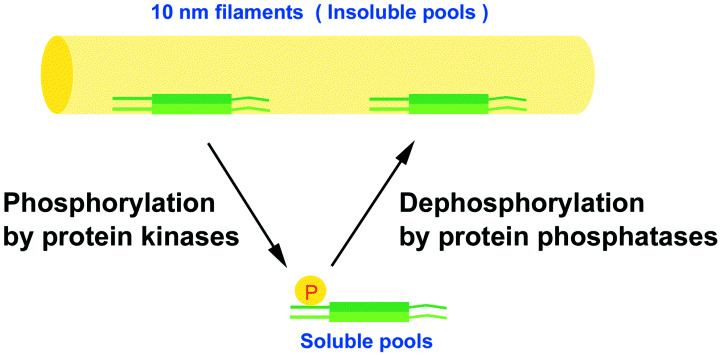
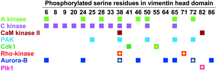
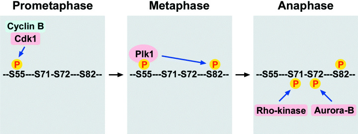
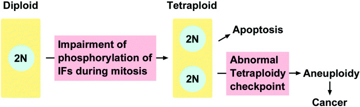
Intermediate filaments (IF) form the structural framework of the cytoskeleton. Although histopathological detection of IF proteins is utilized for examining cancer specimens as reliable markers, the molecular mechanisms by which IF are involved in the biology of cancer cells are still unclear. We found that site-specific phosphorylation of IF proteins induces the disassembly of filament structures. To further dissect the in vivo spatiotemporal dynamics of IF phosphorylation, we developed site- and phosphorylation state-specific antibodies. Using these antibodies, we detected kinase activities that specifically phosphorylate type III IF, including vimentin, glial fibrillary acidic protein and desmin, during mitosis. Cdk1 phosphorylates vimentin-Ser55 from prometaphase to metaphase, leading to the recruitment of Polo-like kinase 1 (Plk1) to vimentin. Upon binding to Phospho-Ser55 of vimentin, Plk1 is activated, and then phosphorylates vimentin-Ser82. During cytokinesis, Rho-kinase and Aurora-B specifically phosphorylate IF at the cleavage furrow. IF phosphorylation by Cdk1, Plk1, Rho-kinase and Aurora-B plays an important role in the local IF breakdown, and is essential for the efficient segregation of IF networks into daughter cells. As another part of our research on IF, we have set out to find the binding partners with simple epithelial keratin 8/18. We identified tumor necrosis factor receptor type 1-associated death domain protein (TRADD) as a keratin 18-binding protein. Together with data from other laboratories, it is proposed that simple epithelial keratins may play a role in modulating the response to some apoptotic signals. Elucidation of the precise molecular functions of IF is expected to improve our understanding of tumor development, invasion and metastasis.
Figures
References
-
- Herrmann H, Hesse M, Reichenzeller M, Aebi U, Magin TM. Functional complexity of intermediate filament cytoskeletons: from structure to assembly to gene ablation. Int Rev Cytol 2003; 223: 83–175. - PubMed
-
- Coulombe PA, Wong P. Cytoplasmic intermediate filaments revealed as dynamic and multipurpose scaffolds. Nat Cell Biol 2004; 6: 699–706. - PubMed
-
- Fuchs E, Cleveland DW. A structural scaffolding of intermediate filaments in health and disease. Science 1998; 279: 514–19. - PubMed
-
- Omary MB, Coulombe PA, McLean WHI. Intermediate filament proteins and their associated diseases. N Engl J Med 2004; 351: 2087–100. - PubMed
-
- Gruenbaum Y, Margalit A, Goldman RD, Shumaker DK, Wilson KL. The nuclear lamina comes of age. Nat Rev Mol Cell Biol 2005; 6: 21–31. - PubMed
Publication types
MeSH terms
Substances
LinkOut - more resources
Full Text Sources
Other Literature Sources
Research Materials
Miscellaneous
