

The role of microorganisms in atopic dermatitis
- PMID: 16542358
- PMCID: PMC1809642
- DOI: 10.1111/j.1365-2249.2005.02980.x
The role of microorganisms in atopic dermatitis
Abstract
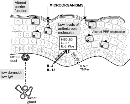
Atopic dermatitis (AD) is a common, fluctuating skin disease that is often associated with atopic conditions such as asthma and IgE-mediated food allergy and whose skin lesions are characterized by a Th-2 cell-mediated response to environmental antigens. The increasing prevalence and severity of atopic diseases including AD over the last three decades has been attributed to decreased exposure to microorganisms during early life, which may result in an altered Th-1/Th-2-balance and/or reduced T cell regulation of the immune response. Patients with AD exhibit defects in innate and acquired immune responses resulting in a heightened susceptibility to bacterial, fungal and viral infections, most notably colonization by S. aureus. Toxins produced by S. aureus exacerbate disease activity by both the induction of toxin-specific IgE and the activation of various cell types including Th-2 cells, eosinophils and keratinocytes. Allergens expressed by the yeast Malazessia furfur, a component of normal skin flora, have also been implicated in disease pathogenesis in a subset of AD patients. Microorganisms play an influential role in AD pathogenesis, interacting with disease susceptibility genes to cause initiation and/or exacerbation of disease activity.
Figures

References
-
- Lee YA, Wahn U, Kehrt R, et al. A major susceptibility locus for atopic dermatitis maps to chromosome 3q21. Nat Genet. 2000;26:470–3. - PubMed
-
- Cookson WO, Ubhi B, Lawrence R, et al. Genetic linkage of childhood atopic dermatitis to psoriasis susceptibility loci. Nat Genet. 2001;27:372–3. - PubMed
-
- Cox HE, Moffatt MF, Faux JA, Walley AJ, Coleman R, Trembath RC, Cookson WO, Harper JI. Association of atopic dermatitis to the beta subunit of the high affinity immunoglobulin E receptor. Br J Dermatol. 1998;138:182–7. - PubMed
-
- He JQ, Chan-Yeung M, Becker AB, Dimwit-Ward H, Ferguson AC, Manfreda J, Watson WT, Sandford AJ. Genetic variants of the IL13 and IL4 genes and atopic diseases in at-risk children. Genes Immun. 2003;4:385–9. - PubMed
Publication types
MeSH terms
Substances
LinkOut - more resources
Full Text Sources
Other Literature Sources
Medical
