. 2006 Mar 28;103(13):4946-51.
doi: 10.1073/pnas.0506352103. Epub 2006 Mar 16.

COMP-angiopoietin-1 promotes wound healing through enhanced angiogenesis, lymphangiogenesis, and blood flow in a diabetic mouse model

Affiliations

COMP-angiopoietin-1 promotes wound healing through enhanced angiogenesis, lymphangiogenesis, and blood flow in a diabetic mouse model

Chung-Hyun Cho et al. Proc Natl Acad Sci U S A. .

Erratum in

  • Proc Natl Acad Sci U S A. 2006 Jun 27;103(26):10146

Abstract

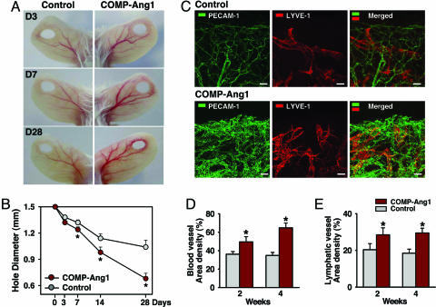
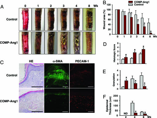
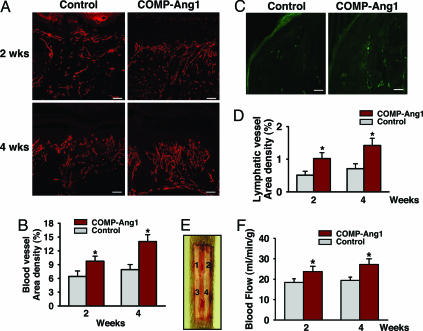
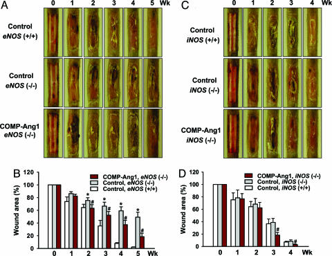
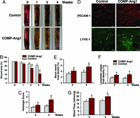
Microvascular dysfunction is a major cause of impaired wound healing seen in diabetic patients. Therefore, reestablishment of structural and functional microvasculature could be beneficial to promote wound healing in these patients. Angiopoietin-1 (Ang1) is a specific growth factor functioning to generate a stable and functional vasculature through the Tie2 and Tie1 receptors. Here we determined the effectiveness of cartilage oligomeric matrix protein (COMP)-Ang1, a soluble, stable, and potent form of Ang1, on promotion of healing in cutaneous wounds of diabetic mice. An excisional full-thickness wound was made in the dorsal side of the tail of diabetic (db/db) mice, and mice were then treated systemically with adenovirus (Ade) encoding COMP-Ang1 or with control virus encoding beta-gal (Ade-beta-gal) or treated topically with recombinant COMP-Ang1 protein or BSA. Time course observations revealed that mice treated with Ade-COMP-Ang1 or COMP-Ang1 protein showed accelerated wound closure and epidermal and dermal regeneration, enhanced angiogenesis and lymphangiogenesis, and higher blood flow in the wound region compared with mice treated with control virus or BSA. COMP-Ang1 promotion of wound closure and angiogenesis was not dependent on endothelial nitric oxide synthase or inducible nitric oxide synthase alone. Taken together, these findings indicate that COMP-Ang1 can promote wound healing in diabetes through enhanced angiogenesis, lymphangiogenesis, and blood flow.


Conflict of interest statement

Conflict of interest statement: No conflicts declared.

Figures

Fig. 1.
Fig. 1.
COMP-Ang1 promotes angiogenesis, lymphangiogenesis, and wound healing in ear skin. FVB/N mice were treated with 1 × 10⁹ pfu of Ade-β-gal (Control) or Ade-COMP-Ang1 (COMP-Ang1) virus, and a closed punched-hole injury was made in the ear. At the indicated days (D) later, ears were photographed (A) and hole diameter was measured (B). (C) Blood and lymphatic vessels at healing margins were visualized with PECAM-1 (green) and LYVE-1 (red) immunostaining, respectively, 28 days after treatment. (Scale bars, 50 μm.) Area densities of blood (D) and lymphatic (E) vessels were measured. Mice treated with COMP-Ang1 show improved wound healing with enhanced densities of blood and lymphatic vessels in ear skin. All circles shown in B and all bars shown in D and E represent mean ± SD from four mice. ∗, P < 0.01 versus control at each time point.
Fig. 2.
Fig. 2.
COMP-Ang1 accelerates wound healing in tail skin of diabetic mice. An excisional full-thickness wound (approximate area, 30 mm2) was made in the tail skin of diabetic db/db mice, and mice were treated with 1 × 10⁹ pfu of Ade-β-gal (Control) or Ade-COMP-Ang1 (COMP-Ang1) virus. At the indicated weeks later, tails were photographed (A), wound areas were measured (B), and regenerative activities of epidermis and dermis (D), granulation thickness (E), and epidermal thickness (F) were measured. ND, not determined. (C) Representative photographs of hematoxylin/eosin (HE) staining and α-smooth muscle actin (α-SMA) (green) and PECAM-1 (red) immunostaining of sections of wound areas of mice treated with COMP-Ang1 and control virus 8 weeks after treatment. (Scale bars, 100 μm.) All bars shown in B and DF represent mean ± SD from five mice. ∗, P < 0.01 versus control at each time point.
Fig. 3.
Fig. 3.
COMP-Ang1 promotes angiogenesis and blood flow in the wound region of tail skin. An excisional full-thickness wound (approximate area, 30 mm2) was made in the tail skin of diabetic db/db mice, and mice were treated with 1 × 10⁹ pfu of Ade-β-gal (Control) or Ade-COMP-Ang1 (COMP-Ang1) virus. Two (A) and four (A and C) weeks later, blood and lymphatic vessels were visualized with PECAM-1 (red) (A) and LYVE-1 (green) (C) immunostaining, and area densities of blood (B) and lymphatic (D) vessels were determined. (Scale bars, 50 μm.) Each bar represents mean ± SD from five mice. (E) By using a laser Doppler flowmeter (Transonic Systems), tissue blood flow in four regions (1, 2, 3, and 4) of the wound area on the dorsal side of the tail were measured. (F) Quantification of skin blood flow at each of the four numbered regions in E was performed, and mean values were obtained 2 and 4 weeks after treatment with control or COMP-Ang1 virus. Each bar represents mean ± SD from four mice. ∗, P < 0.05 versus control at each time point.
Fig. 4.
Fig. 4.
COMP-Ang1 accelerates wound healing in tail skin of eNOS (−/−) and iNOS (−/−) mice. An excisional full-thickness wound (approximate area, 30 mm2) was made in the tail skin of eNOS (+/+), eNOS (−/−), iNOS (+/+), and iNOS (−/−) mice, and mice were treated with 1 × 10⁹ pfu of Ade-β-gal (Control) or Ade-COMP-Ang1 (COMP-Ang1). At the indicated weeks later, tails were photographed (A and C), and wound areas were measured (B and D). Each bar represents the mean ± SD from five mice. ∗, P < 0.05 versus control in eNOS (+/+) at each time point; #, P < 0.05 versus control in eNOS (−/−) or iNOS (−/−) at each time point.
Fig. 5.
Fig. 5.
Topical COMP-Ang1 promotes wound healing with enhanced angiogenesis and blood flow in tail skin. Daily topical treatment with ≈100 μg of BSA (Control) or 100 μg of COMP-Ang1 was applied to part of an excisional full-thickness wound injury (approximate area, 30 mm2) in the tail skin of diabetic db/db mice. At the indicated weeks later, tails were photographed (A) and wound areas and regenerative activities of epidermis and dermis were measured (B and C). Four weeks later, blood and lymphatic vessels were visualized with PECAM-1 (red) or LYVE-1 (green) immunostaining (D). (Scale bars, 50 μm.) At 2 and 4 weeks later, area densities of blood (E) and lymphatic (F) vessels were measured, and blood flow of wound area on the dorsal side of the tail was measured (G) as described in the Fig. 3 legend. Each bar represents mean ± SD from six mice. ∗, P < 0.05 versus control at each time point.

References

    1. Martin P. Science. 1997;276:75–81. - PubMed
    1. Tonnesen M. G., Feng X., Clark R. A. J. Invest. Dermatol. Symp. Proc. 2000;5:40–46. - PubMed
    1. Hirakawa S., Detmar M. J. Dermatol. Sci. 2004;35:1–8. - PubMed
    1. Yancopoulos G. D., Davis S., Gale N. W., Rudge J. S., Wiegand S. J., Holash J. Nature. 2000;407:242–248. - PubMed
    1. Tammela T., Petrova T. V., Alitalo K. Trends Cell Biol. 2005;15:434–441. - PubMed

Publication types

MeSH terms