

The right ventricle in congenital heart disease
- PMID: 16543599
- PMCID: PMC1860730
- DOI: 10.1136/hrt.2005.077438
The right ventricle in congenital heart disease
Abstract
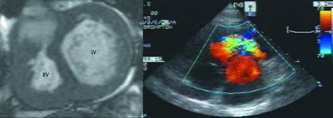
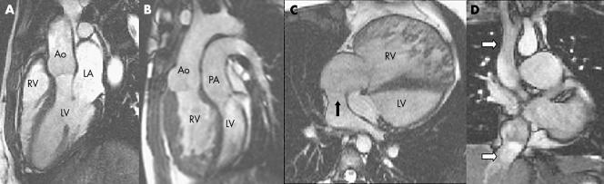
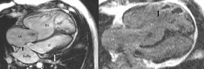
In patients with congenital heart disease the right ventricle (RV) may support the pulmonary (subpulmonary RV) or the systemic circulation (systemic RV). During the last 50 years evidence is accumulating that RV dysfunction develops in many of these patients and leads to considerable morbidity and mortality. Therefore RV function in certain groups of congenital heart disease patients needs close surveillance and timely and appropriate intervention to optimise outcomes. Despite major progress being made, assessing the RV either in the subpulmonary or the systemic circulation remains challenging, often requiring a multi-imaging approach and expertise (echocardiography, magnetic resonance imaging, nuclear and occasionally invasive assessment with angiography). This review discusses the implications of volume and pressure loading of the RV in the context of congenital heart disease and describes the most relevant imaging modalities for monitoring RV function.


References
-
- Samyn M M. A review of the complementary information available with cardiac magnetic resonance imaging and multi‐slice computed tomography (CT) during the study of congenital heart disease. Int J Cardiovasc Imaging 200420569–578. - PubMed
-
- Boxt L M. Magnetic resonance and computed tomographic evaluation of congenital heart disease. J Magn Reson Imaging 200419827–847. - PubMed
-
- Friedberg M K, Rosenthal D N. New developments in echocardiographic methods to assess right ventricular function in congenital heart disease. Curr Opin Cardiol 20052084–88. - PubMed
Publication types
MeSH terms
LinkOut - more resources
Full Text Sources
Other Literature Sources
Medical