. 2006 Apr;116(4):1052-62.
doi: 10.1172/JCI27352. Epub 2006 Mar 16.

Intestinal ABCA1 directly contributes to HDL biogenesis in vivo

Affiliations

Intestinal ABCA1 directly contributes to HDL biogenesis in vivo

Liam R Brunham et al. J Clin Invest. 2006 Apr.

Abstract

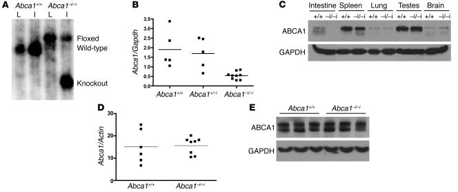
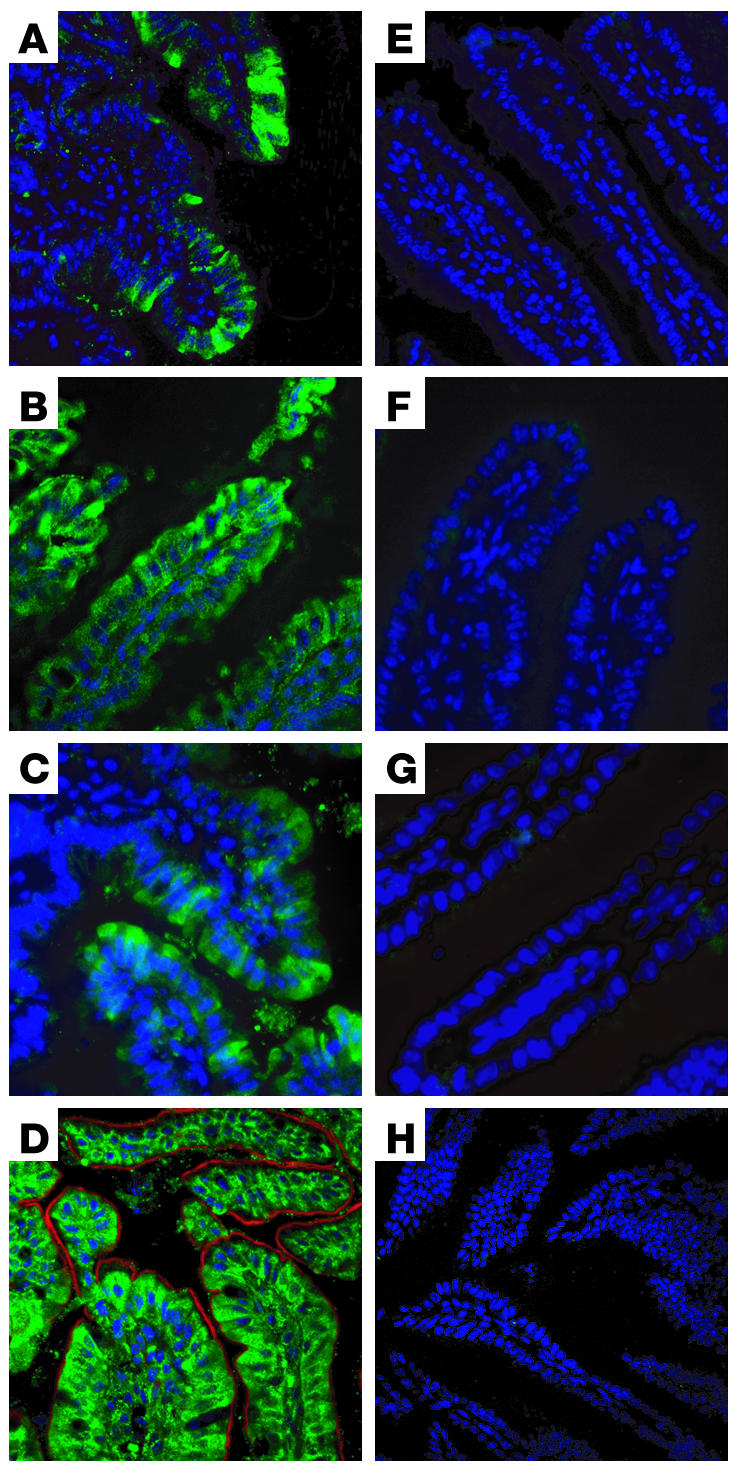
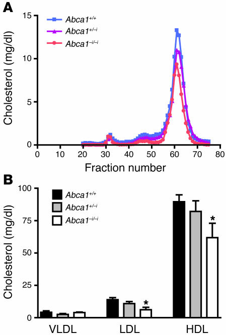
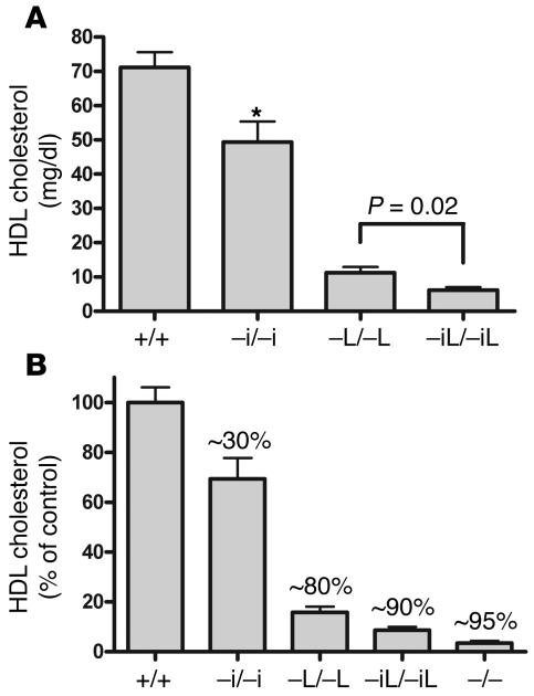
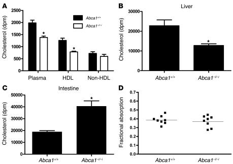
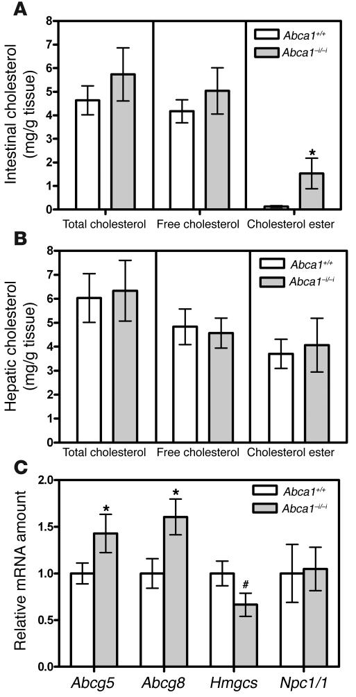
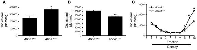
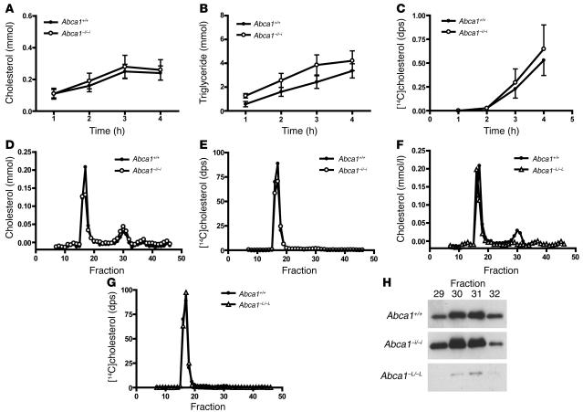
Plasma HDL cholesterol levels are inversely related to risk for atherosclerosis. The ATP-binding cassette, subfamily A, member 1 (ABCA1) mediates the rate-controlling step in HDL particle formation, the assembly of free cholesterol and phospholipids with apoA-I. ABCA1 is expressed in many tissues; however, the physiological functions of ABCA1 in specific tissues and organs are still elusive. The liver is known to be the major source of plasma HDL, but it is likely that there are other important sites of HDL biogenesis. To assess the contribution of intestinal ABCA1 to plasma HDL levels in vivo, we generated mice that specifically lack ABCA1 in the intestine. Our results indicate that approximately 30% of the steady-state plasma HDL pool is contributed by intestinal ABCA1 in mice. In addition, our data suggest that HDL derived from intestinal ABCA1 is secreted directly into the circulation and that HDL in lymph is predominantly derived from the plasma compartment. These data establish a critical role for intestinal ABCA1 in plasma HDL biogenesis in vivo.


Figures

Figure 1
Figure 1. Generation of ABCA1 intestine-specific knockout mice (Abca1–i/–i ).
(A) Southern blot of genomic liver (L) and intestine (I) DNA from mice with WT (+/+) or floxed (–i/–i) alleles in the presence of Cre recombinase. DNA was digested with EcoRV and hybridized with a probe to the genomic region between exons 44 and 45 in the Abca1 gene to produce the 6-kb WT, 7.3-kb floxed, and 4.2-kb knockout bands. (B) Quantitative real-time PCR of RNA isolated from mouse intestine. Reverse-transcribed RNA was amplified with oligos specific for Abca1 and Gapdh. (C) Western blot of tissue lysates from control (+/+) and Abca1–i/–i (–i/–i) mice with antibodies against ABCA1, and GAPDH as loading control. (D) Quantitative real-time PCR of RNA isolated from livers of Abca1+/+and Abca1–i/–i mice. Reverse-transcribed RNA was amplified with oligos specific for Abca1 and Actin. (E) Representative Western blot of liver lysates from Abca1+/+and Abca1–i/–i mice.
Figure 2
Figure 2. Expression of ABCA1 in mouse intestine.
Ten-micron sections of mouse intestine were stained with an antibody against ABCA1 (green), or with DAPI (blue) or phalloidin (red). (AC) Ileum, ×20 magnification (A) and ×40 magnification (B and C), from Abca1+/+ mice. (EG) Corresponding sections from Abca1–i/–i mice. (D) Jejunum, ×60 magnification, from Abca1+/+ mouse. (H) Jejunum, ×60 magnification, from Abca1+/+ mouse stained with random mouse IgG as negative control.
Figure 3
Figure 3. Analysis of plasma lipoproteins by FPLC.
(A) Equal volumes of plasma from Abca1+/+, Abca1+/–i, and Abca1–i/–i mice fasted for 4 hours were pooled and fractionated by FPLC. Total cholesterol in each fraction was determined by enzymatic assay. (B) Plasma from individual mice fasted for 4 hours was fractionated, and cholesterol concentration was determined online by FPLC. n = 6–9 mice per group. *P < 0.05.
Figure 4
Figure 4. Tissue-specific contributions of ABCA1 to plasma HDL cholesterol levels.
(A) Plasma HDL cholesterol levels in control mice, and mice lacking intestinal ABCA1 (–i/–i), hepatic ABCA1 (–L/–L), or both (–iL/–iL). Mice lacking both hepatic and intestinal ABCA1 had a further significant decrease in plasma HDL cholesterol compared with mice lacking hepatic ABCA1. (B) Plasma HDL cholesterol levels as a percentage of those of strain-matched controls. The percentage decrease compared with strain-matched controls is indicated over each bar. Deletion of hepatic and intestinal ABCA1 results in an approximately 90% decrease in plasma HDL cholesterol, similar to that in mice lacking ABCA1 globally (–/–). n ≥ 4 mice per group. *P < .01.
Figure 5
Figure 5. Intestinal cholesterol transport in mice lacking intestinal ABCA1.
(A) Appearance of [3H]cholesterol in whole-plasma, HDL, and non-HDL fractions after 2 hours. Mice were gavaged with 0.2 μCi of [3H]cholesterol, and the appearance of the tracer was assessed after 2 hours. (B) [3H]cholesterol in liver 2 hours after oral gavage. (C) [3H]cholesterol in small intestine 2 hours after oral gavage. The small intestine was rinsed with PBS to remove luminal contents. (D) Fractional cholesterol absorption determined by fecal dual-isotope method. n = 6 mice per group. *P < 0.05.
Figure 6
Figure 6. Tissue cholesterol levels and gene expression.
(A) Intestinal cholesterol levels in control mice and mice lacking intestinal ABCA1. n = 3 mice per group. (B) Hepatic cholesterol levels in control mice and mice lacking intestinal ABCA1. n = 3 mice per group. (C) Relative amounts of various mRNAs in intestines from control mice and mice lacking intestinal ABCA1. Values are relative to the mRNA amount in control mice, which is arbitrarily set as 1. n = 4–8 mice per group. *P < 0.05. #P = 0.06.
Figure 7
Figure 7. Cholesterol secretion from primary enterocytes.
Primary enterocytes from Abca1+/+ and Abca1–i/–i mice were isolated and radiolabeled with [3H]cholesterol, then chased in the presence of micelles. (A) [3H]cholesterol remaining in cells after 2 hours of chase. (B) [3H]cholesterol in media after 2 hours chase. (C) Media were fractionated by ultracentrifugation, and [3H]cholesterol was measured in each fraction. n = 6 per group. *P < 0.05. **P < 0.001.
Figure 8
Figure 8. Analysis of lymph lipoproteins in Abca1+/+ and Abca1–i/–i mice.
(A) Lymph cholesterol transport rate in Abca1+/+ and Abca1–i/–i mice. (B) Lymph triglyceride transport rate in Abca1+/+ and Abca1–i/–i mice. (C) Appearance of [14C]cholesterol in lymph during intraduodenal infusion as a fraction of [3H]oleate. (D) FPLC analysis of lymph from Abca1+/+ and Abca1–i/–i mice. (E) Distribution of [14C]cholesterol in FPLC fractions of Abca1+/+ and Abca1–i/–i mice. (F) FPLC analysis of lymph from Abca1+/+ and Abca1–L/–L mice. (G) Distribution of [14C]cholesterol in FPLC fractions of Abca1+/+ and Abca1–L/–L mice. (H) Western blot of apoA-I in HDL-sized fractions from lymph of Abca1+/+, Abca1–i/–i, and Abca1–L/–L mice. n ≥ 6 mice per group. dps, disintegrations per second.

References

    1. Glomset J.A. The plasma lecithin: cholesterol acyltransferase reaction. J. Lipid Res. 1968;9:155–167. - PubMed
    1. Glomset J.A., Norum K.R. The metabolic role of lecithin: cholesterol acyltransferase. Perspectives from pathology. Adv. Lipid Res. 1973;11:1–65. - PubMed
    1. Linsel-Nitschke P., Tall A.R. HDL as a target in the treatment of atherosclerotic cardiovascular disease. Nat. Rev. Drug Discov. 2005;4:193–205. - PubMed
    1. Francis G.A., Knopp R.H., Oram J.F. Defective removal of cellular cholesterol and phospholipids by apolipoprotein A-I in Tangier disease. J. Clin. Invest. 1995;96:78–87. - PMC - PubMed
    1. Schmitz G., Langmann T. Structure, function and regulation of the ABC1 gene product. Curr. Opin. Lipidol. 2001;12:129–140. - PubMed

Publication types

Substances