Comparative Study
. 2006 Apr;116(4):916-28.
doi: 10.1172/JCI27203.

Yeast zymosan, a stimulus for TLR2 and dectin-1, induces regulatory antigen-presenting cells and immunological tolerance

Affiliations
Comparative Study

Yeast zymosan, a stimulus for TLR2 and dectin-1, induces regulatory antigen-presenting cells and immunological tolerance

Stephanie Dillon et al. J Clin Invest. 2006 Apr.

Abstract

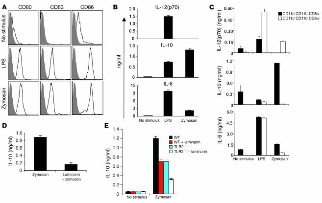
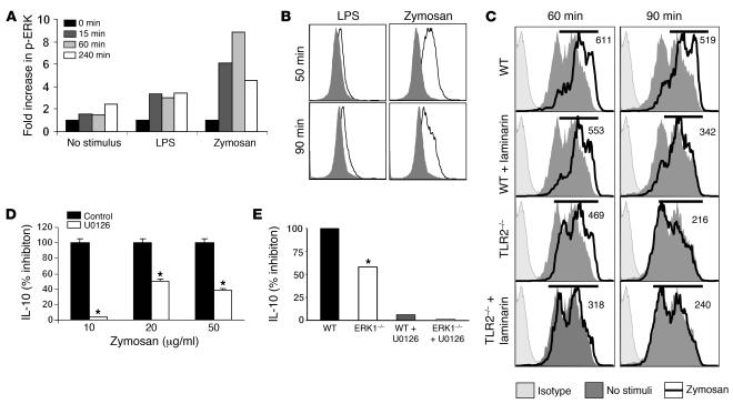
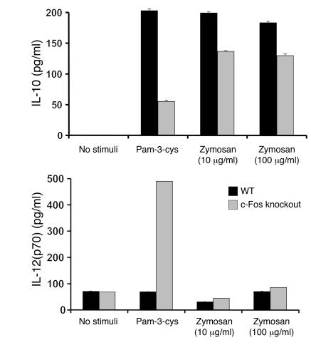
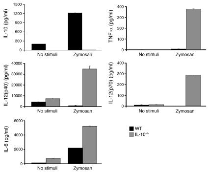
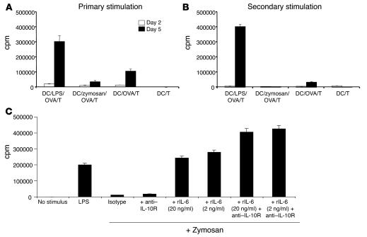
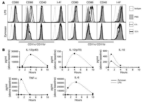
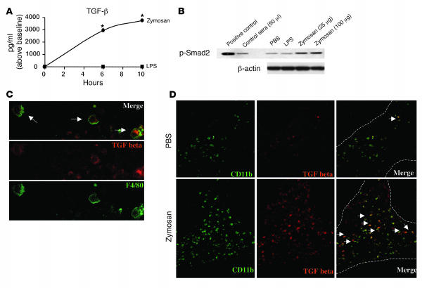
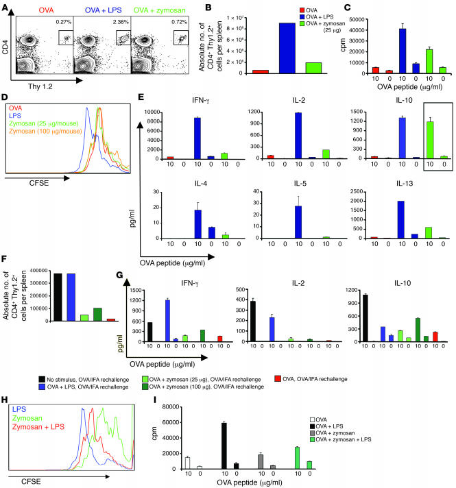
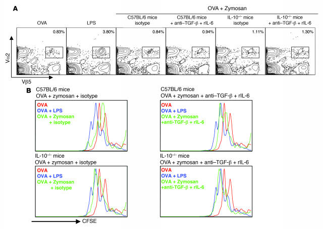
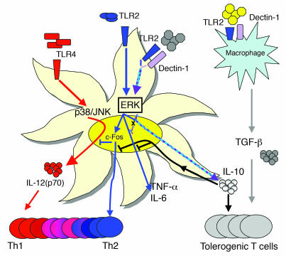
Emerging evidence suggests critical roles for APCs in suppressing immune responses. Here, we show that zymosan, a stimulus for TLR2 and dectin-1, regulates cytokine secretion in DCs and macrophages to induce immunological tolerance. First, zymosan induces DCs to secrete abundant IL-10 but little IL-6 and IL-12(p70). Induction of IL-10 is dependent on TLR2- and dectin-1-mediated activation of ERK MAPK via a mechanism independent of the activation protein 1 (AP-1) transcription factor c-Fos. Such DCs stimulate antigen-specific CD4+ T cells poorly due to IL-10 and the lack of IL-6. Second, zymosan induces F4-80+ macrophages in the splenic red pulp to secrete TGF-beta. Consistent with these effects on APCs, injection of zymosan plus OVA into mice results in OVA-specific T cells that secrete little or no Th1 or Th2 cytokines, but secrete robust levels of IL-10, and are unresponsive to challenge with OVA plus adjuvant. Finally, coinjection of zymosan with OVA plus LPS suppresses the response to OVA via a mechanism dependent on IL-10, TGF-beta, and lack of IL-6. Together, our data demonstrate that zymosan stimulates IL-10+ IL-12(p70)- IL-6low regulatory DCs and TGF-beta+ macrophages to induce immunological tolerance. These data suggest several targets for pharmacological modulation of immune responses in various clinical settings.


Figures

Figure 1
Figure 1. Zymosan induces IL-10 in human and murine DCs via a mechanism involving dectin-1.
(A) Immature, monocyte-derived DCs were cultured for 24–48 hours with E. coli LPS (1 μg/ml) or zymosan (50 μg/ml), and flow cytometric analyses of the expression of the costimulatory molecules CD80 and CD86 and the maturation marker CD83 were performed. Filled histograms show isotype; open histograms represent marker. Data are representative of 10 experiments. (B) Secretion of cytokines in the culture supernatants was measured by ELISA. Data are representative of 18 experiments. (C) Murine, splenic CD11c+CD11b+CD8α, and CD11c+CD11bCD8α+ DC subsets were isolated by flow cytometry and cultured in vitro with E. coli LPS or zymosan for 24–48 hours in the presence of a CD40-L–expressing cell line. Cytokines were analyzed by ELISA. Data are representative of 10 experiments. (D) Immature, monocyte-derived DCs were cultured for 24–48 hours with laminarin (200 μg/ml) plus zymosan or with zymosan alone. IL-10 levels were measured after 24 hours by ELISA. Data are representative of 4 experiments. (E) CD11c+ murine splenic DCs were cultured with laminarin (200 μg/ml) plus zymosan and CD40-L–expressing fibroblasts. IL-10 levels were measured after 24 hours by ELISA. Data are representative of 7 experiments.
Figure 2
Figure 2. Zymosan induces IL-10 in DCs via a mechanism dependent on ERK MAPK.
(A) On day 6, human monocyte-derived DCs were cultured with LPS or zymosan for 0, 15, 60, or 240 minutes. At each time point, the expression of phosphorylated and total ERK was evaluated by ELISA. Data are presented as the fold increases in the phosphorylated to total protein ratios relative to the 0 minute value. Data are representative of 4 experiments. (B) Flow cytometric analyses of phospho-ERK expression in DCs. Filled histograms represent the staining in unstimulated DCs, and the open histograms represent staining after stimulation. Data are representative of 3 experiments. (C) Effect of blocking dectin-1–β-glucan interactions, with laminarin on ERK activation. Data are representative of 3 experiments. Numbers in histograms indicate mean fluorescent intensities of the zymosan-treated DCs. No stimulus indicates DCs cultured in medium alone for 60 or 90 minutes. (D) Effect of blocking ERK1 and 2 with U0126, a synthetic MEK1 and 2 inhibitor, on IL-10 induction by zymosan in human monocyte-derived DCs. Data are representative of 4 experiments. (E) Role of ERK1 and 2 on IL-10 induction by zymosan on CD11c+ murine splenic DCs. DCs from wild-type or ERK1–/– mice were cultured in vitro in the presence of zymosan for 24–48 hours and IL-10 levels assayed by ELISA. Data are representative of 3 experiments. *P < 0.05, control vs. U0126 and WT vs. ERK1–/–.
Figure 3
Figure 3. Zymosan induction of IL-10 in DCs is not dependent on c-Fos.
CD11c+ murine splenic DCs from wild-type or c-Fos–deficient mice were stimulated for 24 hours with zymosan or Pam-3-cys (100 μg/ml) and CD40 ligand cells. IL-10 levels were assayed by ELISA. Data are representative of 3 experiments.
Figure 4
Figure 4. IL-10 regulates the induction of TNF-α, IL-12, and IL-6 in DCs.
Wild-type or IL-10–deficient murine splenic DCs were cultured with zymosan (100 μg/ml) and CD40 ligand cells for 24 hours, and levels of cytokines were assayed by ELISA.
Figure 5
Figure 5. Zymosan-treated DCs induce impaired activation of antigen-specific T cells in vitro via a mechanism dependent on IL-10 and lack of IL-6.
(A) CD11c+ murine splenic DCs (10,000/well) were cultured with naive CD4+CD62L+ OT-II T cells (50,000/well), OVA323–339 peptide (10 μg/ml), and either LPS (1 μg/ml) or zymosan (10 μg/ml) for 2 or 5 days (primary stimulation). (B) T cells were restimulated with plate-bound anti-CD3 and soluble anti-CD28 for an additional 3 days and levels of proliferation determined by incorporation of tritiated thymidine (cpm). (C) Recombinant murine IL-6 (rIL-6) or neutralizing anti–IL-10 receptor (anti–IL-10R) antibodies were added into cultures to determine the role of these cytokines in T cell proliferation induced by zymosan.
Figure 6
Figure 6. Zymosan is not a potent inducer of costimulatory molecules on splenic DCs or proinflammatory cytokines in vivo.
To investigate the effect of zymosan in vivo, C57BL/6 mice were injected with PBS containing either 10 μg E. coli LPS or 25 μg or 100 μg zymosan. (A) Either 4 or 10 hours later, spleens were removed and a small portion digested with collagenase type 4 (1 mg/ml; Worthington Biochemical Corp.) in complete DMEM plus 2% FBS for 30 minutes at 37°C. The red blood cells were lysed and the cell suspension washed twice prior to analysis of cell surface–expressed activation markers by flow cytometry. (B) C57BL/6 mice were injected with PBS containing either 10 μg E. coli LPS or 25 μg zymosan. Blood samples were removed at 1, 4, and 10 hours and serum cytokines analyzed by ELISA.
Figure 7
Figure 7. Zymosan induces TGF-β from spleen macrophages.
(A) C57BL/6 mice were injected i.v. with E. coli LPS (25 μg) or zymosan (25 μg) and bled at various time points; TGF-β induction in the serum was assayed by ELISA. *P < 0.05, zymosan- versus LPS-treated mice. (B) Biological activity of serum TGF-β was determined by stimulating total spleen cells with aliquots of serum and levels of phosphorylation of Smad-2 determined by Western blot. The addition of recombinant TGF-β was used as a positive control. (C) Spleen cells were isolated from the mice used in A at 6 or 10 hours and costained with TGF-β–TRITC and F4-80–FITC, then viewed by confocal microscopy. (D) Immunohistological analysis of TGF-β production. Splenic sections were prepared from spleens isolated at 1, 4, and 10 hours, stained with CD11b–FITC and TGF-β–TRITC, and analyzed by confocal microscopy. Arrows indicate double-positive cells. Original magnification, ×40.
Figure 8
Figure 8. Zymosan induces antigen-specific T cell tolerance in vivo and suppresses the OVA-specific response when coinjected with OVA plus LPS.
(A) B6.PL mice reconstituted with OT-II TCR transgenic T cells were injected i.p. with class II–restricted OVA peptide, OVA323–339 (50 μg) plus LPS (25 μg), OVA323–339 (50 μg) plus zymosan (various doses), or OVA323–339 alone (50 μg). Four days later, spleens were removed and clonal expansion of OVA323–339–specific T cells (A) and numbers of OVA323–329-specific CD4+ T cells per spleen (B) were determined by flow cytometry. (CE) Spleens were isolated 4 days after immunization, and unfractionated spleen cells labeled with CFSE and restimulated in vitro with OVA323–339 for 72 hours. Proliferation as measured by thymidine uptake (C) or CFSE labeling (D) and cytokine production by ELISA (E) were determined. Zymosan induced T cells that produced only high levels of IL-10 (box). Data are representative of 5 independent experiments. (F and G) Mice immunized as above with OVA323–339 plus LPS or OVA323–339 plus zymosan were rechallenged 10 days later with OVA323–339 in IFA. Four days after challenge, clonal expansion and cytokine production upon in vitro restimulation were evaluated. Data are representative of 4 independent experiments. (H and I) B6.PL mice reconstituted with OT-II TCR transgenic T cells were injected with OVA323–339 (50 μg) plus LPS (25 μg), OVA323–339 (50 μg) plus zymosan (100 μg), OVA323–339 (50 μg) plus LPS (25 μg) plus zymosan (100 μg), or OVA323–339 alone (50 μg). Four days later, spleens were removed and unfractionated splenocytes were restimulated with OVA323–339 and (H) CFSE and (I) proliferation of OVA323–339–specific T cells determined.
Figure 9
Figure 9. Impairment of OVA-specific T cell response induced by OVA plus zymosan is dependent on IL-10, TGF-β, and lack of IL-6.
C57BL/6 or IL-10–/– mice were reconstituted with OT-II TCR transgenic T cells on day –1, and the following day, injected with class II–restricted OVA peptide, OVA323–339 (50 μg) plus LPS (25 μg), OVA323–339 (50 μg) plus zymosan (100 μg), or OVA323–339 alone (50 μg). On days –1, 0, and 2, appropriate groups of mice were injected with anti–TGF-β antibody and recombinant murine IL-6. (A) On day 4, clonal expansion was determined by flow cytometry, and (B) splenocytes were isolated, labeled with CFSE, and restimulated in vitro with OVA323–339 to evaluate proliferation of OVA-specific T cells.
Figure 10
Figure 10. A model for induction of regulatory DCs, macrophages, and immune tolerance by yeast zymosan.
Our previous work suggests that Pam-3-cys, which signals DCs through TLR2, induces robust IL-10 and IL-6 but little IL-12(p70) (13, 14). Induction of IL-10 and impaired IL-12(p70) production appear to be critically regulated by ERK activation, which results in the phosphorylation and stabilization of c-Fos, a repressor of IL-12 (13, 14). Such a DC cytokine profile appears to bias toward the Th2 pathway. In the present paper, we demonstrate that zymosan signals to DCs via both TLR2 and dectin-1, inducing the expression of abundant IL-10 but little IL-12(p70) or IL-6. Unlike the case with Pam-3-cys, impairment of IL-12(p70) and induction of c-Fos are not dependent on c-Fos. Induction of IL-10 represses the induction of proinflammatory cytokines, including IL-12, IL-6, and TNF-α. In addition to these effects on DCs, zymosan also induces biologically active TGF-β in macrophages. In this case, the robust induction of IL-10 and TGF-β coupled with impaired induction of IL-6 and IL-12(p70) appears to induce immunological tolerance.

References

    1. Janeway C.A., Jr. Approaching the asymptote? Evolution and revolution in immunology. Cold Spring Harb. Symp. Quant. Biol. 1989;54:1–13. - PubMed
    1. Shortman K., Liu Y.J. Mouse and human dendritic cell subtypes. Nat. Rev. Immunol. 2002;2:151–161. - PubMed
    1. Pulendran B. Modulating vaccine responses with dendritic cells and Toll-like receptors. Immunol. Rev. 2004;199:227–250. - PubMed
    1. Pasare C., Medzhitov R. Toll-like receptors and acquired immunity. Semin. Immunol. 2004;16:23–26. - PubMed
    1. Takeda K., Kaisho T., Akira S. Toll-like receptors. Annu. Rev. Immunol. 2003;21:335–376. - PubMed

Publication types

MeSH terms