. 2006 May;116(5):1346-59.
doi: 10.1172/JCI27414. Epub 2006 Mar 16.

Nephrin ectodomain engagement results in Src kinase activation, nephrin phosphorylation, Nck recruitment, and actin polymerization

Affiliations

Nephrin ectodomain engagement results in Src kinase activation, nephrin phosphorylation, Nck recruitment, and actin polymerization

Rakesh Verma et al. J Clin Invest. 2006 May.

Abstract

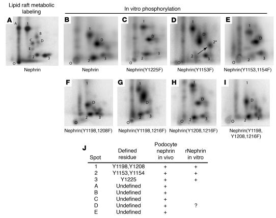
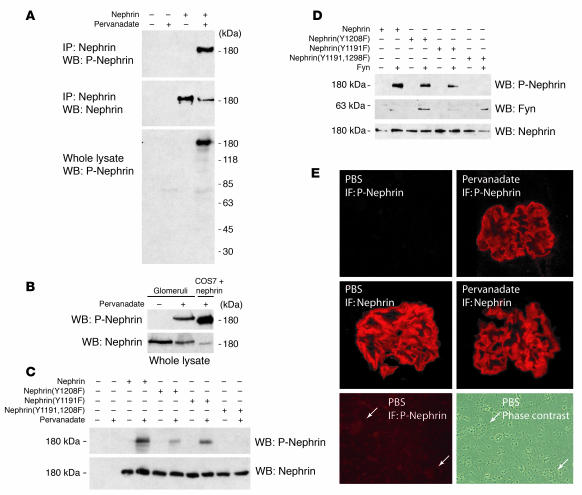
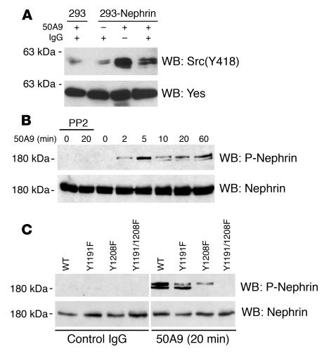
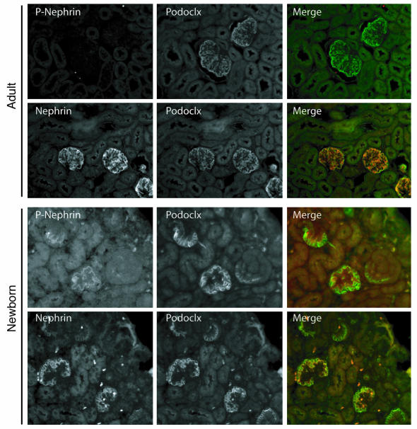
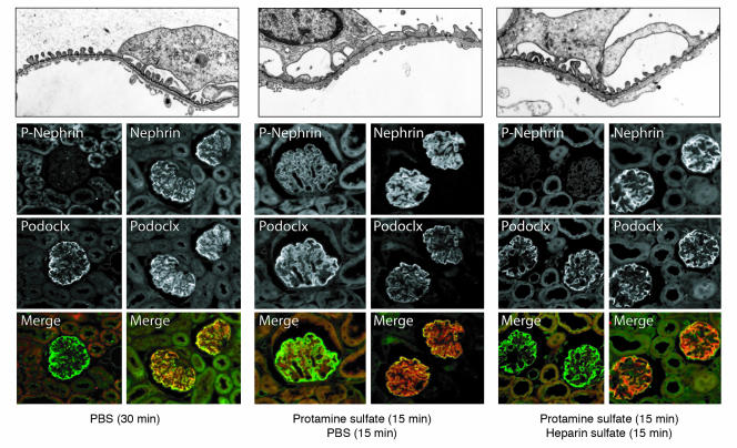
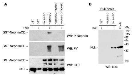
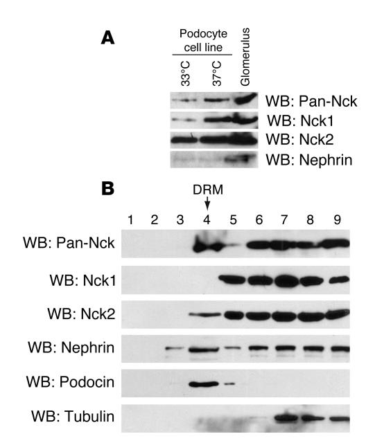
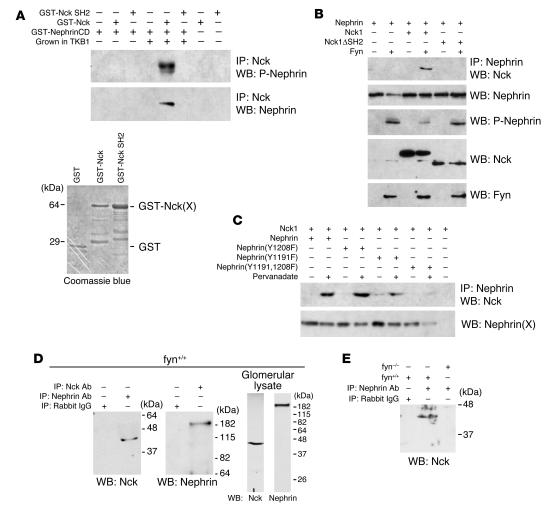
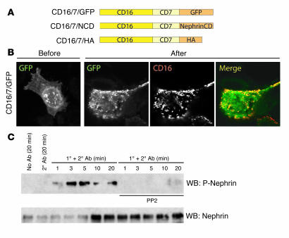
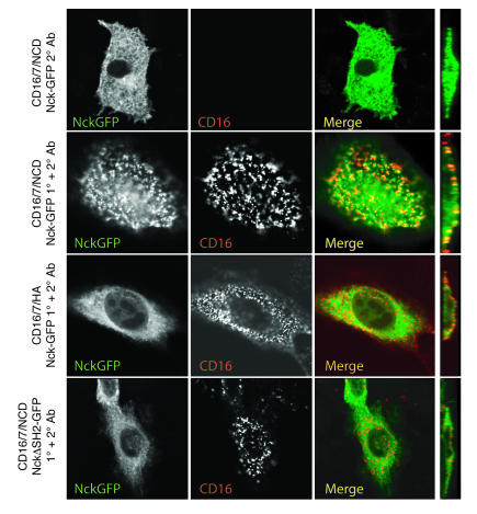
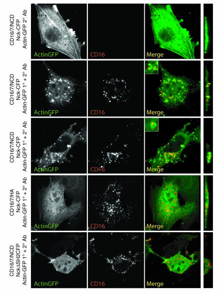
A properly established and maintained podocyte intercellular junction, or slit diaphragm, is a necessary component of the selective permeability barrier of the kidney glomerulus. The observation that mutation or deletion of the slit diaphragm transmembrane protein nephrin results in failure of podocyte foot process morphogenesis and concomitant proteinuria first suggested the hypothesis that nephrin serves as a component of a signaling complex that directly integrates podocyte junctional integrity with cytoskeletal dynamics. The observations made herein provide the first direct evidence to our knowledge for a phosphorylation-mediated signaling mechanism by which this integrative function is derived. Our data support the model that during podocyte intercellular junction formation, engagement of the nephrin ectodomain induces transient Fyn catalytic activity that results in nephrin phosphorylation on specific nephrin cytoplasmic domain tyrosine residues. We found that this nephrin phosphorylation event resulted in recruitment of the SH2-SH3 domain-containing adapter protein Nck and assembly of actin filaments in an Nck-dependent fashion. Considered in the context of the role of nephrin family proteins in other organisms and the integral relationship of actin dynamics and junction formation, these observations establish a function for nephrin in regulating actin cytoskeletal dynamics.


Figures

Figure 1
Figure 1. Identification of Fyn-dependent tyrosine phosphorylation sites on nephrin.
For in vivo metabolic labeling of nephrin (A), DRM obtained from isolated mouse glomeruli was pelleted, resuspended in kinase buffer, and incubated in the presence of [32P-γ]ATP. These DRM were re-extracted in RIPA buffer, and nephrin was obtained thereafter by immunoprecipitation. For in vitro phosphorylation of nephrin by recombinant Fyn (BI), purified recombinant GST-nephrinCD or various GST-nephrinCD point mutants were phosphorylated in vitro by recombinant Fyn. Phosphorylated bands representing nephrin protein or its mutants were separated by SDS-PAGE, identified by autoradiography, and processed for 2D tryptic peptide mapping. Representative tryptic peptide maps for radiophosphate-labeled wild type nephrin obtained by both in vivo (A) and in vitro (B) phosphorylation methods are shown juxtaposed for comparison, and corresponding peptides are labeled as defined in J (along with a summary of peptide characteristics). Mutagenesis of individual tyrosine residues was performed to map Fyn-dependent phosphorylation sites. Where single peptides contained multiple tyrosine residues, single, double, and triple combination tyrosine mutants were created. Shown are representative experiments. Note that for peptide 2, mutation of Y1154 had no effect, Y1153F mutation resulted in a shift in mobility (arrow denotes the shift from 2 to 2*), and mutation of both Y1153 and Y1154 to phenylalanine resulted in disappearance of peptide 2. O, origin; rNephrin, recombinant nephrin.
Figure 2
Figure 2. Characterization of a P-nephrin polyclonal antibody.
(A) Cell lysate — obtained from COS7 cells transfected as labeled with plasmid encoding mouse nephrin and treated with or without pervanadate — was analyzed as indicated by immunoblotting with affinity-purified rabbit polyclonal anti–P-nephrin. (B) Mouse kidney was perfused with pervanadate or PBS; subsequent to glomerular isolation and extraction, glomerular lysate was resolved by SDS-PAGE and immunoblotted for P-nephrin. (C) SYF mouse embryonic fibroblasts were transiently transfected with plasmid encoding nephrin or the indicated nephrin mutants and treated or not treated with pervanadate prior to cell lysis. Cell lysates were resolved by SDS-PAGE and immunoblotted with indicated antibodies. (D) COS7 cells were transiently transfected with plasmids encoding nephrin or the indicated nephrin mutants and with Fyn or a control plasmid. Obtained cell lysates were resolved by SDS-PAGE and immunoblotted with indicated antibodies. (E) Top panels: Adult mouse kidneys were perfused with PBS or pervanadate and were subsequently examined by indirect immunofluorescence (IF) using the indicated primary antibodies. Bottom panels: Indirect immunofluorescence microscopy and phase-contrast microscopy of identical fields was used to confirm absence of P-nephrin staining in glomeruli (see Figure 4). Magnification, ×40 (top 4 panels); ×20 (bottom 2 panels).
Figure 3
Figure 3. Engagement of nephrin extracellular domain results in activation of Src and Fyn-dependent nephrin phosphorylation on Y1191 and Y1208.
HEK 293T cells were stably transfected with plasmids encoding human nephrin or nephrin mutants Y1191F, Y1208F, and Y1191/1208F. Cultured transfectants were stimulated where indicated by addition of mouse anti-human nephrin extracellular domain monoclonal antibody (50A9) or with a mouse IgG control antibody of the same isotype and a secondary anti-mouse IgG. (A) Cells were stimulated for 10 minutes by addition of indicated antibody to culture media and were analyzed by immunoblotting with phosphospecific pan-Src(Y418) antibody or an anti-Yes antibody to demonstrate equivalent loading. (B) Time-course experiment demonstrated induction of nephrin tyrosine phosphorylation upon addition of 50A9 antibody to media. Where indicated, cells were pretreated with PP2 for 15 minutes prior to addition of antibodies. Lysates were immunoblotted with P-nephrin or total nephrin antibodies. (C) Indicated stably transfected 293T cells were stimulated for 10 minutes by addition of indicated antibody to culture media and were analyzed by immunoblotting with the indicated antibodies.
Figure 4
Figure 4. Endogenous nephrin is recognized by P-nephrin antibody in developing but not adult glomeruli.
Newborn or adult mouse kidney sections were prepared as described in Methods. Sections were incubated simultaneously with rabbit P-nephrin or total nephrin antibody and with mouse anti-podocalyxin antibody (Podoclx) and were analyzed by indirect immunofluorescence microscopy. Magnification, ×400.
Figure 5
Figure 5. Nephrin is transiently tyrosine phosphorylated during foot process effacement in the protamine sulfate–induced podocyte injury model.
Adult mouse kidneys were perfused as indicated prior to perfusion fixation with paraformaldehyde. Sections were analyzed by electron microscopy (top row; magnification, ×2,800) or by indirect immunofluorescence microscopy (bottom rows; magnification, ×400) after incubating sections simultaneously with rabbit P-nephrin or total nephrin antibody and with mouse anti-podocalyxin antibody. Protamine sulfate rapidly induced foot process effacement; normal podocyte morphology was nearly restored following perfusion with heparin sulfate. P-nephrin immunoreactivity was detected following protamine sulfate perfusion, but detection was attenuated following perfusion with heparin sulfate.
Figure 6
Figure 6. Nck associates with nephrin.
GST-nephrinCD or its Y1208 mutant was expressed in BL21 or TKB1 E. coli and was affinity purified on glutathione agarose. (A) Immunoblotting with P-nephrin or nonspecific PY antibody demonstrated that expression in TKB1 (but not BL21) cells resulted in phosphorylation on Y1208 and other nephrin tyrosine residues. (B) Purified recombinant GST alone or GST-nephrinCD obtained from either BL21 or TKB1 cells that was bound to glutathione agarose was incubated with isolated mouse glomerular lysate. Washed beads were eluted with glutathione. Eluate was resolved by SDS-PAGE and immunoblotted with the panel of antibodies shown in Table 2. Representative immunoblot was obtained using a pan-Nck antibody that demonstrated association of Nck with tyrosine-phosphorylated wild-type GST-nephrinCD but not unphosphorylated GST-nephrinCD or GST alone.
Figure 7
Figure 7. Nck1 and Nck2 are present in podocytes.
(A) Lysate obtained from an immortalized podocyte cell line cultured under permissive (33°C) or nonpermissive (37°C) conditions or obtained from isolated mouse glomeruli was resolved by SDS-PAGE and immunoblotted using the indicated antibodies. (B) Mouse glomeruli isolated by sieving were extracted in buffer containing 1% Triton X-100 at 4°C. The lysate was subjected to flotation gradient centrifugation. DRM was identified in lane 5 at the interface between 5 and 30% Optiprep densities. Fractions (1 ml) were collected and analyzed by immunoblotting with the indicated antibodies.
Figure 8
Figure 8. Nck binds directly with nephrin via an SH2 domain–dependent interaction.
(A) Purified recombinant GST-nephrinCD was prepared from BL21 cells or TKB1 cells and was incubated in vitro with GST-Nck or a Nck mutant in the SH2 domain. The specified mixtures of recombinant proteins were immunoprecipitated with anti-Nck. Immune complexes were resolved by SDS-PAGE and were immunoblotted with either nephrin or P-nephrin antibody. Coomassie blue–stained gel demonstrates GST-Nck proteins used in this experiment. (B) COS7 cells were transiently transfected as indicated with plasmids encoding nephrin and Fyn. Independently, COS7 cells were transfected with plasmid encoding Nck1 or Nck1ΔSH2. Lysate obtained from these independently transfected cells were mixed, then immunoprecipitated and/or immunoblotted as indicated. (C) COS7 cells were transfected with plasmid encoding nephrin or its tyrosine mutants, then treated with pervanadate for 15 minutes as indicated prior to lysis. Independently, COS7 cells were transfected with plasmid encoding Nck1. Lysate obtained from these independently transfected cells were mixed, then immunoprecipitated and/or immunoblotted as indicated. (D) Coimmunoprecipitation experiments from isolated wild-type mouse glomerular lysate using pan-Nck and nephrin antibodies demonstrating association of endogenous Nck and nephrin. (E) Similar experiments performed on isolated glomerular lysate obtained from wild-type and Fyn-null mice.
Figure 9
Figure 9. Clustering of CD16 chimeric proteins and induction of tyrosine phosphorylation on clustered CD16/7/nephrinCD (CD16/7/NCD).
(A) Schematic representation of CD16 fusion proteins prepared for these experiments. (B) Confocal microscopy imaging of NIH 3T3 cells expressing the CD16/7/GFP fusion protein before and after addition to media of live cells of anti-CD16 antibody and rhodamine-conjugated anti-IgG antibody. In the merged image at right, rhodamine IgG (red) and GFP (green) colocalized (yellow) on the plasma membrane, demonstrating clustering of the CD16/7/GFP chimeric protein. Note absence of clusters where anti-CD16 antibody was not present. Magnification, ×600. (C) NIH 3T3 cells expressing CD16/7/nephrinCD were treated with clustering antibodies (1° + 2° Ab) or as indicated for the time periods shown, then immunoblotted with P-nephrin antibody to detect phosphorylation of Y1191 and Y1208. Pretreatment of cells with PP2 blocked tyrosine phosphorylation on these sites following induction of clusters.
Figure 10
Figure 10. Nck is recruited to CD16/7/nephrinCD clusters at the plasma membrane.
NIH 3T3 cells expressing indicated chimeric proteins (red) and Nck-GFP or NckΔSH2-GFP (green) were treated with anti-CD16 antibody and rhodamine anti-IgG antibody or were treated with the secondary antibody only, then fixed and examined by confocal microscopy. Far right panels are images reconstructed in the YZ plane. Colocalization of Nck-GFP and CD16/7/nephrinCD appear yellow on merged images. Data are representative of multiple experiments. Magnification, ×600.
Figure 11
Figure 11. Clustered nephrinCD induces Nck-dependent actin polymerization.
NIH 3T3 cells expressing the indicated chimeric proteins (red), Nck-CFP or NckΔSH2-CFP, and actin-GFP (green) were treated with anti-CD16 antibody and rhodamine anti-IgG antibody or were treated with the secondary antibody only, then fixed and examined by confocal microscopy. Far right panels are images reconstructed in the YZ plane. Two representative examples of changes that occur in the presence of actin-GFP, Nck-CFP, and CD16/7/nephrinCD are shown in the second and third rows — note the intense green actin-GFP signal emitted by multiple actin tails or pedestals was associated nearly invariably with the red signal that marked clustered CD16/7/nephrinCD at the plasma membrane (insets). Data are representative of multiple experiments. Magnification, ×600; ×1,200 (insets).

Comment in

Similar articles

Cited by

References

    1. Kerjaschki D. Caught flat-footed: podocyte damage and the molecular bases of focal glomerulosclerosis. J. Clin. Invest. 2001;108:1583–1587. doi: 10.1172/JCI200114629. - DOI - PMC - PubMed
    1. Johnstone D., Holzman L.B. Clinical impact of research on the podocyte slit diaphragm. Nature Clinical Practice Nephrology. 2006 In press. - PubMed
    1. Kestila M., et al. Positionally cloned gene for a novel glomerular protein--nephrin--is mutated in congenital nephrotic syndrome. Mol. Cell. 1998;1:575–582. - PubMed
    1. Putaala H., Soininen R., Kilpelainen P., Wartiovaara J., Tryggvason K. The murine nephrin gene is specifically expressed in kidney, brain and pancreas: inactivation of the gene leads to massive proteinuria and neonatal death. Hum. Mol. Genet. 2001;10:1–8. - PubMed
    1. Hamano Y., et al. Determinants of vascular permeability in the kidney glomerulus. J. Biol. Chem. 2002;277:31154–31162. - PubMed

Publication types