. 2006;38(8):1417-28.
doi: 10.1016/j.biocel.2006.02.003. Epub 2006 Mar 3.

Nucleocapsid protein of SARS-CoV activates the expression of cyclooxygenase-2 by binding directly to regulatory elements for nuclear factor-kappa B and CCAAT/enhancer binding protein

Affiliations

Nucleocapsid protein of SARS-CoV activates the expression of cyclooxygenase-2 by binding directly to regulatory elements for nuclear factor-kappa B and CCAAT/enhancer binding protein

Xiaohong Yan et al. Int J Biochem Cell Biol. 2006.

Retraction in

Abstract

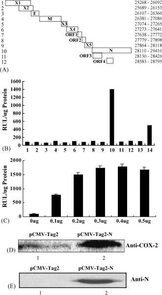
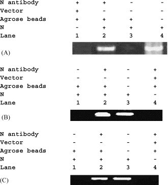
SARS-associated coronavirus (SARS-CoV) causes inflammation and damage to the lungs resulting in severe acute respiratory syndrome. To evaluate the molecular mechanisms behind this event, we investigated the roles of SARS-CoV proteins in regulation of the proinflammatory factor, cyclooxygenase-2 (COX-2). Individual viral proteins were tested for their abilities to regulate COX-2 gene expression. Results showed that the COX-2 promoter was activated by the nucleocapsid (N) protein in a concentration-dependent manner. Western blot analysis indicated that N protein was sufficient to stimulate the production of COX-2 protein in mammalian cells. COX-2 promoter mutations suggested that activation of COX-2 transcription depended on two regulatory elements, a nuclear factor-kappa B (NF-kappaB) binding site, and a CCAAT/enhancer binding protein (C/EBP) binding site. Electrophoretic mobility shift assay (EMSA) and chromatin immunoprecipitation (ChIP) demonstrated that SARS-CoV N protein bound directly to these regulatory sequences. Protein mutation analysis revealed that a Lys-rich motif of N protein acted as a nuclear localization signal and was essential for the activation of COX-2. In addition, a Leu-rich motif was found to be required for the N protein function. A sequence of 68 residuals was identified as a potential DNA-binding domain essential for activating COX-2 expression. We propose that SARS-CoV N protein causes inflammation of the lungs by activating COX-2 gene expression by binding directly to the promoter resulting in inflammation through multiple COX-2 signaling cascades.


Figures

Fig. 1
Fig. 1
SARS-CoV N protein activates the expression of COX-2 in 293T cells. (A) Diagram of individual open reading frames of SARS-CoV with locations on the viral genome. (B) Analysis COX-2 promoter activation by individual SARS-CoV proteins. 293T cells were cotransfected with plasmid expressing different SARS-CoV proteins, respectively, and the reporter plasmid in which the luciferase gene is under the control of COX-2 promoter. Relative luciferase activity was determined by standard procedures. The expressed genes are indicated in panel A: 13 is the negative control pCMV-Tag2 and 14 is the positive control pCMV-Tag2-HBx (X protein of hepatitis B virus). (C) Analysis of dose-dependent of the SARS-CoV N protein in the activation of COX-2 promoter. 293T cells were cotransfected with different amount of plasmids expressing N protein along with the reporter plasmid and relative luciferase activity was determined. Values correspond to an average of at least three independent experiments done in duplicate. Error bars show 1 S.D. (D) Western blot analysis of the expression of COX-2 protein activated by N protein. Cells were transfected with empty vector pCMV-Tag2 as a control (lane 1) or with plasmid pCMV-Tag-N, expressing SARS-CoV N protein (lane 2). Cell extracts were prepared and the expressed proteins were determined using rabbit anti-COX-2 antibody. (E) Western blot analysis of same blot probed with antibodies against N protein.
Fig. 2
Fig. 2
Functional analysis of cis-regulatory elements of the COX-2 promoter. Diagram of individual cis-regulatory elements of the COX-2 promoter and its truncated or site-specific mutants are shown in the left panel and results from luciferase activity assay are shown in the right panel. Plasmid carrying the SARS-CoV N gene and plasmids containing the luciferase reporter gene driven by individual COX-2 promoter mutants were cotransfected into 293T cells. Promoter activities were determined by measuring the relative luciferase activity in transfected-cell lysates. pCMV-Tag2 was used as a vector only control. Luciferase activities correspond to an average of at least three independent experiments done in duplicate. The black symbols indicate mutations. Error bars show 1 S.D.
Fig. 3
Fig. 3
Determination of interaction between SARS-CoV N protein and COX-2 promoter by electrophoretic mobility shift assay (EMSA). EMSA was performed with nuclear extracts of 293T cells transfected with (lanes 4–6) or without (lanes 1–3) the N gene. Probes were generated by annealing single-stranded and end-labeled oligonucleotides containing the cognate COX-2 promoter regions. C/EBP at nucleotides −132/−125 (A) or NF-κB at nucleotides −228/−204 (B) probes were added to all reactions (lanes 1–6). Unlabeled double-stranded oligonucleotide competitors were added during preincubation prior to probe addition (lanes 1 and 5). For supershift experiments, polyclonal antibody was incubated with nuclear extracts before adding to the reaction (lane 6). Samples were electrophoresed on 5% nondenaturing polyacrylamide gel and visualized by autoradiography. Arrows indicate the super shifted protein–DNA complexes.
Fig. 4
Fig. 4
Determination of interaction between SARS-CoV N protein and COX-2 promoter by chromatin immunoprecipitation (ChIP) assays. 293T cells transfected with empty vector pCMV-Tag2 (lane 1 in A; lane 4 in B and C) or with pCMV-Tag2-N expressing the N protein (lanes 2–4 in A; lanes 1–3 in B; lanes 1–3 in C) were lysed and subjected to ChIP assays. Immunoprecipitated complexes were collected, subjected to PCR amplification, and separated by agarose gel electrophoresis. (A) The COX-2 promoter region (−502 to −2) was amplified by PCR using specific primers (CP1 and CP2). (B) The COX-2 promoter region (−243 to −136) amplified by PCR using NF-κB-specific primers (NF-κB-CP1 and NF-κB-CP2). (C) The COX-2 promoter region (−155 to −2) amplified by PCR using C/EBP-specific primers (C/EBP-CP1 and CP2).
Fig. 5
Fig. 5
Functional analysis of SARS-CoV N protein by sequential deletions. (A) Diagram of N-terminal truncated mutants of the SARS-CoV N protein and determination of functions of N-terminal truncated N proteins by measuring their ability on the activation of the COX-2 promoter. (B) Diagram of C-terminal truncated mutants of the SARS-CoV N protein and determination of functions of C-terminal truncated N proteins by measuring their ability on the activation of the COX-2 promoter. 293T cells were cotransfected with plasmids containing individual mutant N gene expressing different truncated protein and the reporter gene. The effects of each mutant protein on the activation of COX-2 promoter were measured by the luciferase activity assay. pCMV-Tag2 was used as a vector control. Values correspond to an average of at least three independent experiments done in duplicate. Error bars show 1 S.D.
Fig. 6
Fig. 6
Determination of the function of putative nuclear localization signals of the N protein. (A) Diagram and location of the potential nuclear localization signals of the N protein and their deletion mutants. MutN1 with 38-PKQRRPQ-44 deleted; MutN2 with 220-LALLLLDRLNRL-231 deleted; MutN3 with 257-KKPRQKR-263 deleted; MutN4 with 369-KKDKKKK-376 deleted. (B) Functional analysis of deletion mutant N proteins. 293T cells were cotransfected with plasmids carrying genes expressing the mutant N proteins and the reporter vector. The effects of the deletions on the N protein were determined by measuring luciferase activity. pCMV-Tag2 was used as a vector control. Values correspond to an average of at least three independent experiments done in duplicate. Error bars show 1 S.D. (C) Western bolt analysis of N proteins expressed in transfected cells using N protein antibody.

References

    1. Bagetta G., Corasaniti M.T., Paoletti A.M., Berliocchi L., Nistico R., Giammarioli A.M. HIV-1 gp120-induced apoptosis in the rat neocortex involves enhanced expression of cyclooxygeanse type2 (COX-2) Biochem. Biophys. Res. Commun. 1998;244:819–824. - PubMed
    1. He R., Dobie F., Ballantime M., Leeson A., Li Y., Bastien N. Analysis of multimerization of the SARS coronavirus nucleocapsid protein. Biochem. Biophys. Res. Commun. 2004;317:1030–1036. - PMC - PubMed
    1. He R., Leeson A., Andonov A., Li Y., Bastien N., Cao J. Activation of AP-1 signal transduction pathway by SARS coronavirus nucleocapsid protein. Biochem. Biophys. Res. Commun. 2003;311:870–876. - PMC - PubMed
    1. Kin Y., Fischer S.M. Transcriptional regulation of cyclooxygenase-2 in mouse skin carcinoma cells. J. Biol. Chem. 1998;273:27686–27694. - PubMed
    1. Ksiazek T.G., Erdman D., Goldsmith C.S., Zaki S.R., Peret T., Emery S. A novel coronavirus associated with severe acute respiratory syndrome. N. Engl. J. Med. 2003;348:1953–1966. - PubMed

Publication types

MeSH terms