. 2006 Apr;188(7):2325-35.
doi: 10.1128/JB.188.7.2325-2335.2006.

Phenotypic characterization of Streptococcus pneumoniae biofilm development

Affiliations

Phenotypic characterization of Streptococcus pneumoniae biofilm development

Magee Allegrucci et al. J Bacteriol. 2006 Apr.

Abstract

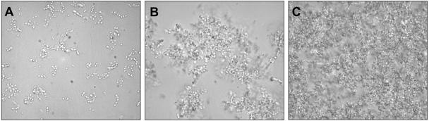
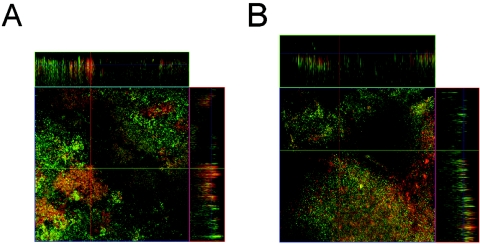
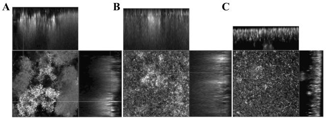
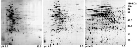
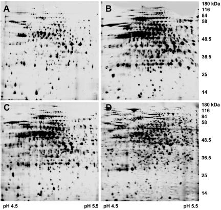
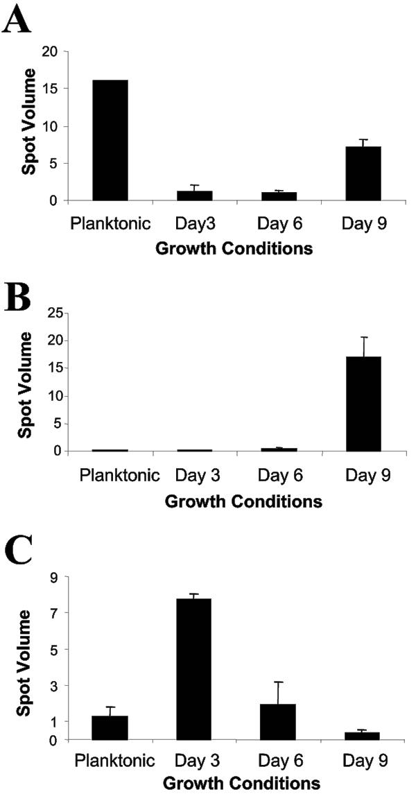
Streptococcus pneumoniae is among the most common pathogens associated with chronic otitis media with effusion, which has been hypothesized to be a biofilm disease. S. pneumoniae has been shown to form biofilms, however, little is known about the developmental process, the architecture, and the changes that occur upon biofilm development. In the current study we made use of a continuous-culture biofilm system to characterize biofilm development of 14 different S. pneumoniae strains representing at least 10 unique serotypes. The biofilm development process was found to occur in three distinct stages, including initial attachment, cluster formation, and biofilm maturation. While all 14 pneumococcal strains displayed similar developmental stages, the mature biofilm architecture differed significantly among the serotypes tested. Overall, three biofilm architectural groups were detected based on biomass, biofilm thickness, and cluster size. The biofilm viable cell counts and total protein concentration increased steadily over the course of biofilm development, reaching approximately 8 x 10(8) cells and approximately 15 mg of protein per biofilm after 9 days of biofilm growth. Proteomic analysis confirmed the presence of distinct biofilm developmental stages by the detection of multiple phenotypes over the course of biofilm development. The biofilm development process was found to correlate not only with differential production of proteins but also with a dramatic increase in the number of detectable proteins, indicating that biofilm formation by S. pneumoniae may be a far more complex process than previously anticipated. Protein identification revealed that proteins involved in virulence, adhesion, and resistance were more abundant under biofilm growth conditions. A possible role of the identified proteins in biofilm formation is discussed.


Figures

FIG. 1.
FIG. 1.
Streptococcus pneumoniae biofilm formation under continuous flow. Phase-contrast micrographs at a magnification of 1,000× obtained 1 day (A), 2 days (B), and 3 days (C) after initiation of S. pneumoniae biofilm formation. Biofilms were viewed using an Olympus BX60 microscope. Flow-cell experiments were performed in triplicate as described in Materials and Methods.
FIG. 2.
FIG. 2.
Confocal laser scanning microscopic images of (A) 6-day and (B) 9-day-old Streptococcus pneumoniae serotype 3 biofilms. Biofilms were grown in flow cells under once-through flow conditions for 6 and 9 days, after which time the biofilms were stained with the Live/Dead BacLight stain. Biofilms were viewed at 400× magnification. Flow cell experiments were performed in triplicate as described in Materials and Methods.
FIG. 3.
FIG. 3.
Confocal laser scanning microscopic images of 6-day-old biofilms of Streptococcus pneumoniae serotypes. Representative CLSM images of S. pneumoniae group I biofilm architecture (A) of strain BS71 (serotype 3), group II biofilm architecture (B) of strain BS73 (serotype 6), and group III biofilm architecture (C) of strain BS75 (serotype 19) are shown. Biofilms were grown in flow cells under once-through flow conditions for 6 days, after which time the biofilms were stained with the Live/Dead BacLight stain. Biofilms were viewed at 400× magnification. The CLSM images show the xy and xz planes. Flow cell experiments were performed in triplicate as described in Materials and Methods.
FIG. 4.
FIG. 4.
2D images of total cell extracts obtained from 6-day-old Streptococcus pneumoniae capsule serotype 3 biofilms. The total protein extracts (200 μg) were separated using (A) pH 3 to 10NL, (B) pH 4 to 7 and (C) pH 4.5 to 5.5 Immobiline Dry Strips in the first dimension, followed by SDS-PAGE using 11% polyacrylamide gels in the second dimension. Gels were stained with silver nitrate (10).
FIG. 5.
FIG. 5.
2D images of total cell extracts obtained from Streptococcus pneumoniae serotype 3 grown planktonically (A) and as 3-day (B), 6-day (C), and 9-day (D) biofilm. The crude protein extracts (200 μg) were separated on pH 4.5 to 5.5 Immobiline Dry strips, followed by SDS-PAGE using 11% polyacrylamide gels. Gels were stained with silver nitrate (10). Experiments were performed in triplicate.
FIG. 6.
FIG. 6.
Protein production patterns of selected proteins over the course of Streptococcus pneumoniae serotype 3 biofilm development. Representative production patterns of glucose-6-phosphate isomerase (A), glycl-tRNA synthetase (B), and pneumolysin (C). The production pattern analysis was carried out from 2D images that were scanned using a calibrated image scanner (GE Healthcare). Image analysis was done using the Image Master 2D Platinum software (GE Healthcare). Computational analysis was based on 2D protein spot volume. The planktonic growth stage and three biofilm developmental stages were analyzed thus: planktonic, planktonic cell stage; 3 days, 3-day-old biofilms; 6 days, 6-day-old biofilms, and 9 days, 9-day-old biofilms. The data shown here represent the average spot volumes. Experiments were carried out in triplicate for each growth stage.

References

    1. Applegate, D. H., and J. D. Bryers. 1991. Effects on carbon and oxygen limitations and calcium concentrations on biofilm removal processes. Biotechnol. Bioeng. 37:17-25. - PubMed
    1. Arrecubieta, C., R. Lopez, and E. Garcia. 1994. Molecular Characterization of cap3A, a gene from the operon required for the synthesis of the capsule of Streptococcus pneumoniae type 3: sequencing of mutations responsible for the unencapsulated phenotype and localization of the capsular cluster on the pneumococcal chromosome. J. Bacteriol. 176:6375-6383. - PMC - PubMed
    1. Austrian, R., and J. Gold. 1964. Pneumococcal bacteremia with especial reference to bacteremic pneumococcal pneumonia. Ann. Intern. Med. 60:759-776. - PubMed
    1. Balachandran, P., S. K. Hollingshead, J. C. Paton, and D. E. Briles. 2001. The autolytic enzyme LytA of Streptococcus pneumoniae is not responsible for releasing pneumolysin. J. Bacteriol. 183:3108-3116. - PMC - PubMed
    1. Bennett, K. E., M. P. Haggard, P. A. Silva, and I. A. Stewart. 2001. Behaviour and developmental effects of otitis media with effusion into the teens. Arch. Dis. Child. 85:91-95. - PMC - PubMed

Publication types

MeSH terms

Substances

LinkOut - more resources