. 2006 May;173(1):131-49.
doi: 10.1534/genetics.106.055749. Epub 2006 Mar 17.

Searching for neuronal left/right asymmetry: genomewide analysis of nematode receptor-type guanylyl cyclases

Affiliations

Searching for neuronal left/right asymmetry: genomewide analysis of nematode receptor-type guanylyl cyclases

Christopher O Ortiz et al. Genetics. 2006 May.

Abstract

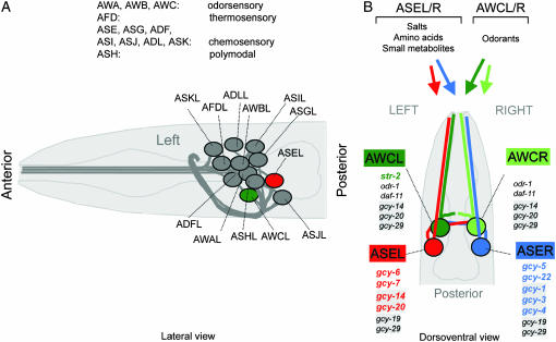
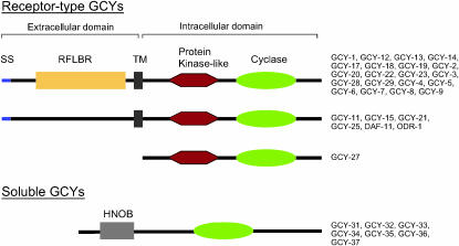
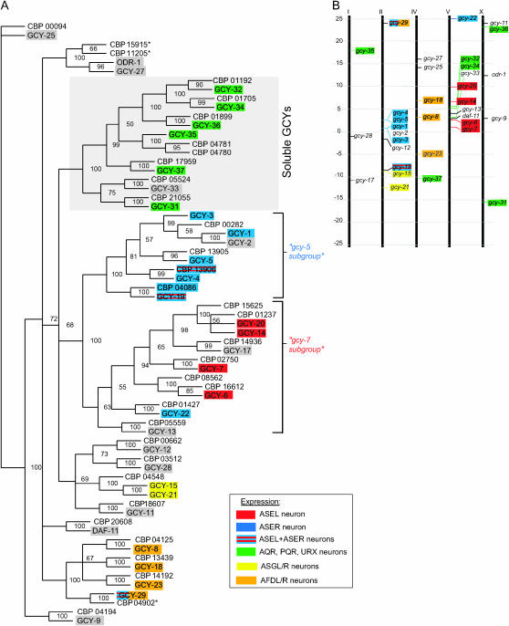
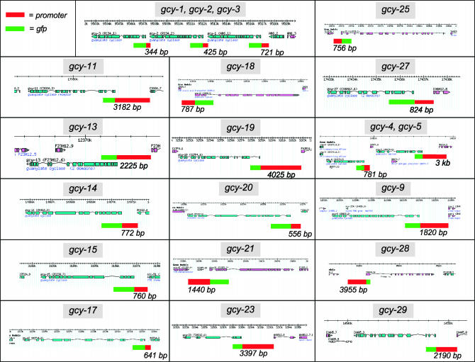
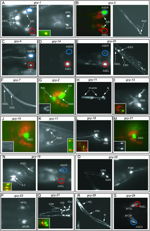
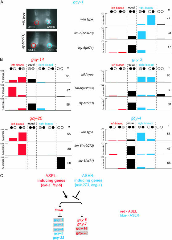
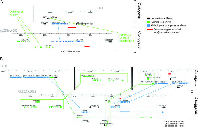
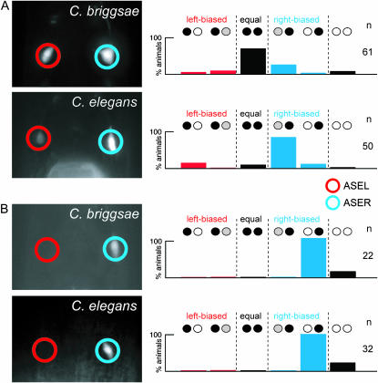
Functional left/right asymmetry ("laterality") is a fundamental feature of many nervous systems, but only very few molecular correlates to functional laterality are known. At least two classes of chemosensory neurons in the nematode Caenorhabditis elegans are functionally lateralized. The gustatory neurons ASE left (ASEL) and ASE right (ASER) are two bilaterally symmetric neurons that sense distinct chemosensory cues and express a distinct set of four known chemoreceptors of the guanylyl cyclase (gcy) gene family. To examine the extent of lateralization of gcy gene expression patterns in the ASE neurons, we have undertaken a genomewide analysis of all gcy genes. We report the existence of a total of 27 gcy genes encoding receptor-type guanylyl cyclases and of 7 gcy genes encoding soluble guanylyl cyclases in the complete genome sequence of C. elegans. We describe the expression pattern of all previously uncharacterized receptor-type guanylyl cyclases and find them to be highly biased but not exclusively restricted to the nervous system. We find that >41% (11/27) of all receptor-type guanylyl cyclases are expressed in the ASE gustatory neurons and that one-third of all gcy genes (9/27) are expressed in a lateral, left/right asymmetric manner in the ASE neurons. The expression of all laterally expressed gcy genes is under the control of a gene regulatory network composed of several transcription factors and miRNAs. The complement of gcy genes in the related nematode C. briggsae differs from C. elegans as evidenced by differences in chromosomal localization, number of gcy genes, and expression patterns. Differences in gcy expression patterns in the ASE neurons of C. briggsae arise from a difference in cis-regulatory elements and trans-acting factors that control ASE laterality. In sum, our results indicate the existence of a surprising multitude of putative chemoreceptors in the gustatory ASE neurons and suggest the existence of a substantial degree of laterality in gustatory signaling mechanisms in nematodes.


Figures

Figure 1.
Figure 1.
An introduction to C. elegans sensory anatomy. (A) A prominent and well-characterized subset of C. elegans sensory neurons, the amphid sensory neurons. As with most other neuron classes, amphid sensory neuron classes consist of one pair of two bilaterally symmetric cells (see also B). Each of the 12 pairs of amphid sensory neurons extends a dendrite to the tip of the nose and an axon into the nerve ring, a nerve bundle where synaptic connections are made (White et al. 1986). Delineated functions of amphid sensory neurons are indicated (Bargmann and Mori 1997). (B) Amphid sensory neuron classes consist of two bilaterally symmetric pairs of neurons, two of which, AWCL/R and ASEL/R, are functionally lateralized. While some other amphid sensory neurons appear to contribute to gustation, ASE is the main gustatory neuron class in C. elegans, mediating responses to salts, amino acids, and small metabolites (Bargmann and Horvitz 1991). ASE mediates not only attractive, but also repulsive, responses to specific chemicals (Sambongi et al. 1999). The salts sodium, chloride, and potassium are sensed in a left/right asymmetric manner, with ASEL sensing sodium, but not chloride and potassium, and ASER sensing chloride and potassium (Pierce-Shimomura et al. 2001). It is not yet known whether other ASE-sensed chemicals may also activate ASEL and ASER differentially. ASE-expressed gcy genes are shown; those newly described in this article are shaded; those that are asymmetric are colored. The AWCL/R neurons can discriminate benzaldehyde and butanone on the basis of the left/right asymmetric expression of str-2 (colored), a G-protein coupled receptor, which is stochastically expressed in either AWCL or AWCR (Troemel et al. 1999; Wes and Bargmann 2001). Newly identified gcy genes in AWC are shaded.
Figure 2.
Figure 2.
Domain structure of GCY proteins. SS, signal sequence; TM, transmembrane domain; RFLBR, receptor family ligand-binding region (PF01094); HNOB, heme nitric oxide-binding domain (PF07700); protein kinase-like, protein kinase domain (PF00069); cyclase, adenylate and guanylate cyclase catalytic domain (PF00211). See supplemental Figure 1 and supplemental Figure 2 at http://www.genetics.org/supplemental/ for the primary sequence alignment of individual domains. We note that several of the receptor-type GCY proteins, such as GCY-11, lack a clear SS at the N terminus but the presence of a clear TM and/or RFLBR domain make us suspect that the absence is due to an incorrectly predicted N terminus of the respective genes and we therefore grouped these genes together with other clear-cut SS/TM-containing proteins. GCY-22 has an unusual and phylogenetically conserved insertion between the transmembrane and protein kinase domain (supplemental Figure 2 at http://www.genetics.org/supplemental/).
Figure 3.
Figure 3.
Sequence similarity and chromosomal localization of gcy genes. (A) Phylogenetic tree based on the intracellular domain of the receptor-type guanylyl cyclases and the complete sequences of the soluble guanylyl cyclases. Numbers at the tree nodes are bootstrap values, which indicate the frequency (in percentages) of occurrence of a given partition in the 1000 replicate trees. C. elegans proteins are shaded; C. briggsae proteins all carry the prefix “CBP.” A select number of cells that coexpress multiple gcy genes are indicated by color-coded shading, as indicated in the inset. See materials and methods for comments on the gcy gene names. Note that the CBP15915*, CBP11205*, and CBP04902* proteins used here differ from those in Wormbase WS149 on the basis of an alternative gene prediction that we performed (see materials and methods). (B) Chromosomal localization of gcy genes. See Table 2 for detailed map position. Chromosome III does not contain any gcy genes.
Figure 4.
Figure 4.
Reporter gene constructs. Representation of genomic loci are adapted from http://www.wormbase.org. Reporter gene constructs are indicated by the 5′ upstream region used (red box), usually the intergenic region up to the next gene, and the gfp coding region (green box; not drawn to scale). See Table 1 for primer sequences and a list of transgenic arrays containing the individual reporter gene constructs.
Figure 5.
Figure 5.
Expression patterns of gcy reporter gene fusions. Transgenic animals expressing gfp reporter gene fusions are shown. Images are of representative animals from several independent lines (see Table 1 for a list of transgenic reporter arrays used). Most transgenic animals are scored in the late larval and adult stage and contain otIs151 in the background to facilitate the identification of the ASE neurons (see materials and methods); blue circles indicate ASER; red circles indicate ASEL. The quantification of the left/right asymmetric expression in ASE is shown in Figure 6. (A) gcy-1prom∷gfp. Dorsal view (left) and ventral view (right) of two different focal plains of the head region. The inset in the right panel shows the overlap of the gfp signal with otIs133, an AIY-expressed rfp marker (lateral view). (B) gcy-3prom∷gfp. Dorsal view of the head region (left) of an animal whose amphid sensory neurons have been filled with DiI. (Right) A full-length worm with expression in the PVT interneuron. (C) gcy-4prom∷gfp. Dorsal view of the head region. (D) gcy-14prom∷gfp. Dorsal view of the head region. (E) gcy-20prom∷gfp. Dorsal view of the head region (left). A full-length worm with expression in the excretory system (right). EXG, excretory gland cell; EXC, excretory canal cell. (F) gcy-7prom∷gfp. Lateral view. Expression in ASEL, but not expression in the excretory canal cell (EXC), has been previously reported (Yu et al. 1997). (G) gcy-2prom∷gfp. Lateral view of the head region. A defined subset of the amphid neurons are filled with DiI to allow for easier assessment of cell position. (Inset) A dorsal view illustrating bilateral symmetry of gfp-expressing cells. (H) gcy-11prom∷gfp. Lateral view of the head region. The strong neuronal expression (N) in the pharynx is likely due to the injection marker, but pharyngeal muscle expression is due to the reporter gene. (I) gcy-13prom∷gfp. Dorsal view of the head region. (Inset) A lateral view of a DiI-filled animal. (J) gcy-15prom∷gfp. Lateral view of the head region. A defined subset of the amphid neurons are filled with DiI to allow for easier assessment of cell position. (Inset) A ventral view to illustrate bilateral symmetry. (K) gcy-17prom∷gfp. Dorsal view of the tail region. (Inset) A DiI-filled animal. (L) gcy-18prom∷gfp. Lateral view of the head region. (Inset) A lateral view of gfp expression in relation to the rfp expression from otIs133, an AIY-specific cell marker. (M) gcy-21prom∷gfp. Lateral view of the head region of a DiI-filled animal. (Inset) A ventral view illustrating bilateral symmetry. (N) gcy-19prom∷gfp. Lateral view (left) and dorsal view (right) of the head region. (Inset, left) The overlap of the gfp signal with DiI-filled IL2 neurons. (O) gcy-25prom∷gfp. (Left) An oblique view of the head region. (Right) A lateral view of the tail region. (P) gcy-23prom∷gfp. Ventral view of the head region. (Q) gcy-27prom∷gfp. Dorsal view of the head region. (Inset) A lateral view of a DiI-filled animal. (R) gcy-28prom∷gfp. VNC, ventral nerve cord; HG, head ganglia; N, non-neuronal cells. The animal is mosaic and does not show muscle expression. (S) gcy-29prom∷gfp. Ventral view of the head region. The reporter is also expressed in AWCL/R and in AVKL/R (not shown in this animal).
Figure 6.
Figure 6.
Regulation of the expression of asymmetric gcy genes. (A and B) Quantification of the asymmetry of ASE-expressed gcy genes in wild-type and mutant backgrounds. (A) Quantification and a representative example of gcy-1prom∷gfp reporter gene-carrying animals. (B) Quantification of the results obtained with the other asymmetrically expressed and previously uncharacterized gcy reporter gene constructs. Black, white, and gray circles indicate relative expression levels of gfp in the ASEL and ASER neurons. Data shown are for one representative array each (otEx2419 for gcy-1prom∷gfp, otEx2423 for gcy-3prom∷gfp, otEx2409 for gcy-4prom∷gfp, otEx2322 for gcy-14prom∷gfp, and otEx2327 for gcy-20prom∷gfp). These arrays were each crossed into the indicated mutant backgrounds. Comparable results were obtained with several independent arrays (not shown). As in other figures, red indicates ASEL expression and blue indicates ASER expression. (C) Summary of the gene regulatory interactions. Gray shading indicates genes identified in this article. For more details on the ASEL and ASER inducers, see Johnston et al. (2005).
Figure 7.
Figure 7.
Functional analysis of gcy-5. (A) Mutant alleles of gcy-5. Color coding for the domains encoded by the individual exons is shown in Figure 2. Reading frames are indicated to illustrate the effects of the respective deletion alleles. tm897 contains a 691-bp deletion from position 462–1045 of the coding sequence and replacement with 5′-GGGGTAGAAGAGGC. Within the genomic locus, the deletion starts in exon 4 and ends in exon 7. With the deletion and insertion, a frameshift is created, leading to an early stop codon. This allele is therefore a putative null allele. The effect of the other alleles is more difficult to predict since the respective deletions start in exons and end in introns. If one assumes splicing around the half-deleted exons, then the two ok alleles produce large, but in-frame, deletions. (B) Chemotaxis to soluble ions of wild-type and gcy-5 mutant animals. NaCl measures the functionality of both ASEL and ASER. NH4Cl mainly measures ASER function since NH4+ is sensed in a ASE-independent manner [NH4+ sensation is unaffected in che-1 mutants in which ASEL/R fails to develop (Chang et al. 2004)]. All strains were grown and assayed at room temperature (21°–23°). Population chemotaxis assays were performed in a radial gradient of the indicated salt (see materials and methods). Each experiment was done with at least two plates in parallel and for each assay plate at least 20 worms reached either attractant or negative control spot. Three to nine independent experiments were done for each condition. Error bars indicate standard error of the mean. For statistical analysis, a one-way ANOVA was performed for each attractant, with Dunnet's post-test comparing all three alleles to wild-type control data. None of the means were significantly different.
Figure 8.
Figure 8.
Analysis of synteny of gcy genes in C. elegans and C. briggsae. Gene predictions were taken from WormBase release WS149 (http://www.wormbase.org). The red line indicates the genomic regions included in gfp reporter gene constructs. The C. elegans reporter constructs are also shown in Figure 4. The size of the C. briggsae gcy-19 construct is 2 kb; the size of the C. briggsae gcy-4 construct is 433 bp (up to the preceding gene).
Figure 9.
Figure 9.
Phylogenetic conservation of gcy gene expression profiles. C. briggsae reporter gene constructs are shown in Figure 8; C. elegans reporter gene constructs are shown in Figure 4. Species names in pictures indicate into which species the respective reporter gene was injected. (A) gcy-4 expression. C. briggsae gcy-4prom∷gfp (CBP13906) is expressed in both ASEL and ASER in C. briggsae (three lines; data for one representative array, otEx2508, are shown), but is expressed predominantly in ASER in C. elegans (three lines; n > 40 each; data for one representative array, otEx2510, are shown). Expression of C. elegans gcy-4prom∷gfp (construct in Figure 4) in C. elegans is also biased to ASER (Figure 5C; Figure 6B). (B) gcy-19 expression. C. briggsae gcy-19prom∷gfp (CBP04086) is expressed exclusively in ASER in C. briggsae (three lines; data for one representative array, otEx2139, are shown) and in C. elegans (three lines; data for one representative array, otEx2141, are shown).

Similar articles

Cited by

References

    1. Bargmann, C. I., 1998. Neurobiology of the Caenorhabditis elegans genome. Science 282: 2028–2033. - PubMed
    1. Bargmann, C. I., and H. R. Horvitz, 1991. Chemosensory neurons with overlapping functions direct chemotaxis to multiple chemicals in C. elegans. Neuron 7: 729–742. - PubMed
    1. Bargmann, C. I., and I. Mori, 1997. Chemotaxis and thermotaxis, pp. 717–738 in C.elegans II, edited by D. L. Riddle, T. Blumenthal, B. J. Meyer and J. R. Priess. Cold Spring Harbor Laboratory Press, Cold Spring Harbor, NY. - PubMed
    1. Bigelow, H. R., A. S. Wenick, A. Wong and O. Hobert, 2004. CisOrtho: a program pipeline for genome-wide identification of transcription factor target genes using phylogenetic footprinting. BMC Bioinformatics 5: 27. - PMC - PubMed
    1. Birnby, D. A., E. M. Link, J. J. Vowels, H. Tian, P. L. Colacurcio et al., 2000. A transmembrane guanylyl cyclase (DAF-11) and Hsp90 (DAF-21) regulate a common set of chemosensory behaviors in Caenorhabditis elegans. Genetics 155: 85–104. - PMC - PubMed

Publication types

MeSH terms

Substances

LinkOut - more resources