. 2006 Mar 28;103(13):5179-84.
doi: 10.1073/pnas.0507175103. Epub 2006 Mar 17.

Essential role for hematopoietic prostaglandin D2 synthase in the control of delayed type hypersensitivity

Affiliations

Essential role for hematopoietic prostaglandin D2 synthase in the control of delayed type hypersensitivity

Seema G Trivedi et al. Proc Natl Acad Sci U S A. .

Abstract

Hematopoietic prostaglandin D(2) synthase (hPGD(2)S) metabolizes cyclooxygenase-derived prostaglandin (PG) H(2) to PGD(2), which is dehydrated to cyclopentenone PGs, including 15-deoxy-Delta(12,14)-PGJ(2) (15d-PGJ(2)). PGD(2) acts through two receptors (DP1 and DP2/CRTH2), whereas 15d-PGJ(2) can activate peroxisome proliferator-activated receptors or inhibit a range of proinflammatory signaling pathways, including NF-kappaB. Despite eliciting asthmatic and allergic reactions through the generation of PGD(2), it is not known what role hPGD(2)S plays in T helper (Th)1-driven adaptive immunity. To investigate this question, the severity and duration of a delayed type hypersensitivity reaction was examined in hPGD(2)S knockout and transgenic mice. Compared with their respective controls, knockouts displayed a more severe inflammatory response that failed to resolve, characterized histologically as persistent acute inflammation, whereas transgenic mice had little detectable inflammation. Lymphocytes isolated from inguinal lymph nodes of hPGD(2)S(-/-) animals showed hyperproliferation and increased IL-2 synthesis effects that were rescued by 15d-PGJ(2), but not PGD(2), working through either of its receptors. Crucially, 15d-PGJ(2) exerted its suppressive effects through the inhibition of NF-kappaB activation and not through peroxisome proliferator-activated receptor signaling. In contrast, lymph node cultures from transgenics proliferated more slowly and synthesized significantly less IL-2 than controls. Therefore, contrary to its role in driving Th2-like responses, this report shows that hPGD(2)S may act as an internal braking signal essential for bringing about the resolution of Th1-driven delayed type hypersensitivity reactions. Consequently, hPGD(2)S-derived cyclopentenone PGs may protect against inflammatory diseases, where T lymphocytes play a pathogenic role, as in rheumatoid arthritis, atopic eczema, and chronic rejection.


Conflict of interest statement

Conflict of interest statement: No conflicts declared.

Figures
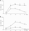
Fig. 1.
Fig. 1.
hPGD2S controls the severity and duration of DTH responses. C57BL/6 mice lacking hPGD2S (■) and wild-type controls (□) (A) along with FVB mice overexpressing this enzyme (♦) and their appropriate controls (□) (B) were sensitized at the base of the tail and challenged in the left paw 14 d later with mBSA, which induced a DTH reaction. Contralateral paws received saline. Paw swelling measurements were made by water-displacement plethysmometry at selected time points after challenge. n = 6–8 animals per group; ∗, P ≤ 0.05; ∗∗, P ≤ 0.01, as determined by ANOVA, followed by Bonferroni t test, with data expressed as mean ± SEM.
Fig. 2.
Fig. 2.
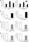
hPGD2S acts as a braking signal for lymphocyte proliferation and IL-2 synthesis. Inguinal lymph nodes from hPGD2S−/− mice previously sensitized to mBSA to trigger a DTH reaction were incubated with the specific antigen (mBSA) for 72 h (A), revealing significantly increased proliferation of these cells, and for 24 h (B), showing enhanced IL-2 synthesis. Similar experiments were carried out on mice overexpressing hPGD2S, where proliferation (C) and IL-2 synthesis (D) were found to be significantly reduced compared with controls. n = 5 animals per group; ∗, P ≤ 0.05; ∗∗, P ≤ 0.01, as determined by t test, with data expressed as mean ± SEM.
Fig. 3.
Fig. 3.
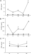
Synthesis of hPGD2S-derived PGD2 and 15d-PGJ2 increases during inflammatory resolution. Inguinal lymph nodes were excised from groups of mBSA-sensitized wild-type and hPGD2S−/− mice 8, 24, 48, and 96 h after mBSA challenge and cultured for 24 h to allow for accumulation of PGs within the culture medium. Synthesis of PGD2 (A) and its metabolite 15d-PGJ2 (B) was found to increase during resolution in wild-type (□) animals, with levels barely detectable in hPGD2S−/− (■) samples. (C) PGE2 was also measured in culture supernatants and found not to change substantially through the time course of the reaction in wild types (□), and no significant difference was detectable in PGE2 levels between wild types and knockouts (■). n = 3–5 animals per group. Data are expressed as mean ± SEM.
Fig. 4.
Fig. 4.
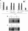
The 15d-PGJ2, but not PGD2, reverses lymphocyte hyperproliferation and IL-2 synthesis in hPGD2S−/− mice. Inguinal lymph nodes were taken from mBSA-sensitized animals 24 h after in vivo challenge and incubated ex vivo for 24 h with BW245C (DP1 receptor), 15R-PGD2 (DP2 receptor agonist), and 15d-PGJ2. Only 15d-PGJ2 was found to reverse the hyperproliferative phenotype (A) and IL-2 synthesis (B) of cells deficient in hPGD2S, whereas DP1 and DP2 agonists were without effect. In the experiments shown in B, BW245C and 15R-PGD2 were also used at 30 nM and 2 μM, respectively, without detectable effect (data not shown). Experiments with a second DP2 receptor agonist, 15(R)-15 methyl PGD2 at 1.25, 2.5, and 5.0 nM confirmed original findings (data not shown). Importantly, the neutralizing property of 15d-PGJ2 was effective at doses that did not cause lymphocyte apoptosis. n = 3–5 animals per group; ∗, P ≤ 0.05; ∗∗, P ≤ 0.01, as determined by ANOVA, followed by post hoc Dunnett’s test, with data expressed as mean ± SEM
Fig. 5.
Fig. 5.
The inhibitory property of 15d-PGJ2 on lymphocyte proliferation is not mediated through PPAR signaling but, at least in part, through inhibition of NF-κB. (A) Inguinal lymph nodes were taken from mBSA-sensitized hPGD2S−/− and wild-type animals 24 h after in vivo challenge and incubated for 24 h with a PPARγ agonist (rosiglitazone) or antagonist (GW9662) as well as 15d-PGJ2 alone or with GW9662, where GW9662 was preincubated for 1 h before adding the cyPG. As before (Fig. 4), only 15d-PGJ2 altered proliferation, whereas PPARγ agonists and antagonists were without effect. Even prior blocking of PPARγ did not alter the rescuing effects of 15d-PGJ2 on knockout cells when compared with corresponding controls. A PPARα agonist (GW7647, 0.1 and 1.0 μg/ml) as well as a PPARβ/δ agonist (L-165041, 0.01–1 μM) were also without effect on proliferation (data not shown). In contrast, NF-κB binding was found to be greatly enhanced in lymph node extract from hPGD2S−/− animals (B), including freshly isolated cells from sensitized mice (labeled C) as well as those stimulated for 24 h with either the specific antigen (mBSA, labeled M) or Con A (nonspecific, labeled A). The converse also held true for hPGD2S transgenic mice (C), where NF-κB binding was reduced in all treatment groups. n = 3–5 animals per group; ∗∗, P ≤ 0.01, as determined by ANOVA, followed by post hoc Dunnett’s test, with data expressed as mean ± SEM.

References

    1. Flower R. J. Pharmacol. Rev. 1974;26:33–67. - PubMed
    1. Gilroy D. W., Lawrence T., Perretti M., Rossi A. G. Nat. Rev. Drug Discov. 2004;3:401–416. - PubMed
    1. Serhan C. N. Histochem. Cell Biol. 2004;122:305–321. - PubMed
    1. Gilroy D. W., Colville-Nash P. R., Willis D., Chivers J., Paul-Clark M. J., Willoughby D. A. Nat. Med. 1999;5:698–701. - PubMed
    1. Luster A. D., Tager A. M. Nat. Rev. Immunol. 2004;4:711–724. - PubMed

Publication types

MeSH terms