

Development of atrophy of the retinal pigment epithelium around disciform scars
- PMID: 16547324
- PMCID: PMC1857011
- DOI: 10.1136/bjo.2005.083022
Development of atrophy of the retinal pigment epithelium around disciform scars
Abstract
Background/aims: Eyes with burnt out disciform scars secondary to age related macular degeneration (AMD) are regarded as visually stable. The aim of this study is to report the subsequent development of atrophy of the retinal pigment epithelium (RPE) around the scars and discuss the possible basis.
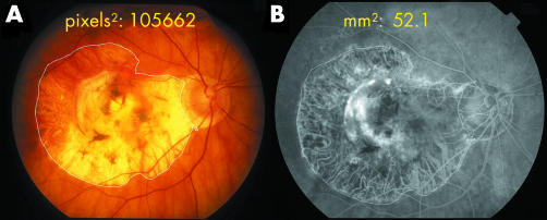
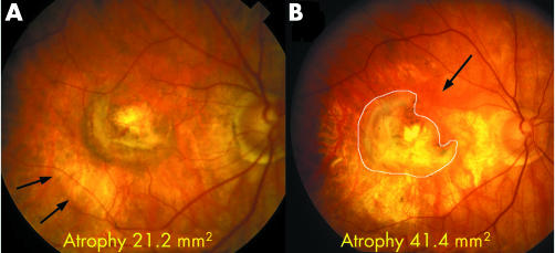
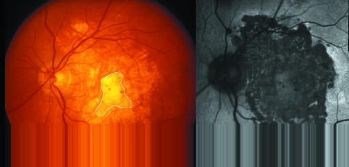
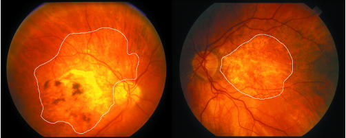
Methods: 20 eyes from 18 patients were observed to develop atrophy around choroidal neovascularisation (CNV). A method of measuring expansion of the atrophy over time is described using the Topcon Imagenet 2000 system. An additional 10 clinicopathological examples were reviewed.
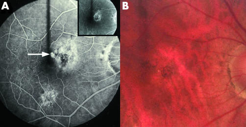
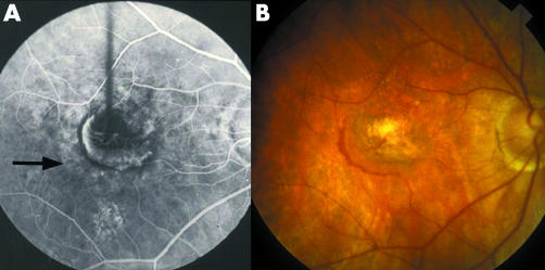
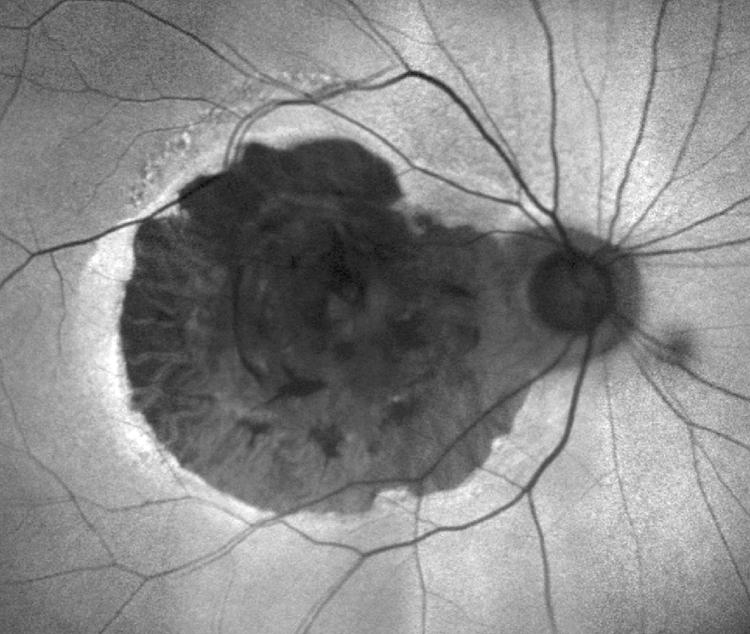
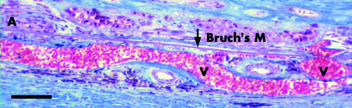
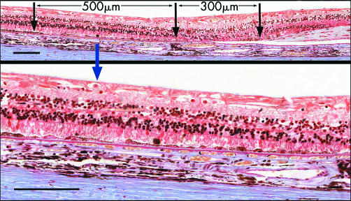
Results: Clinically CNV became surrounded initially by a ring of pallor that progressed to an expanding band of atrophy of the RPE. It developed most rapidly in the first 3 years after CNV became quiescent but then continued to expand slowly to more than three times the size of the scar. Histopathological specimens showed large choroidal vessels entering the scars directly and a reduced number of small choroidal vessels beneath and around the scar
Conclusions: Disciform scars may become surrounded by an expanding band of atrophy of the RPE, postulated to result from remodelling of the choroidal circulation. The ongoing enlargement of the resulting scotoma may need to be considered when planning management and assessing treatment outcomes.
Comment in
-
Progressive RPE atrophy around disciform scars.Br J Ophthalmol. 2006 Apr;90(4):396-7. doi: 10.1136/bjo.2005.086041. Br J Ophthalmol. 2006. PMID: 16547308 Free PMC article.
-
Choroidal neovascularisation and atrophy.Br J Ophthalmol. 2006 Apr;90(4):398-9. doi: 10.1136/bjo.2005.084830. Br J Ophthalmol. 2006. PMID: 16547309 Free PMC article.
Similar articles
-
Progressive RPE atrophy around disciform scars.Br J Ophthalmol. 2006 Apr;90(4):396-7. doi: 10.1136/bjo.2005.086041. Br J Ophthalmol. 2006. PMID: 16547308 Free PMC article.
-
Risk factors for choroidal neovascularization and geographic atrophy in the complications of age-related macular degeneration prevention trial.Ophthalmology. 2008 Sep;115(9):1474-9, 1479.e1-6. doi: 10.1016/j.ophtha.2008.03.008. Epub 2008 May 27. Ophthalmology. 2008. PMID: 18502512 Clinical Trial.
-
The development of choroidal neovascularization in eyes with the geographic atrophy form of age-related macular degeneration.Ophthalmology. 1999 May;106(5):910-9. doi: 10.1016/S0161-6420(99)00509-6. Ophthalmology. 1999. PMID: 10328389
-
Retinal pigment epithelial tear following photodynamic therapy for choroidal neovascularization secondary to AMD.Eye (Lond). 2005 Dec;19(12):1315-24. doi: 10.1038/sj.eye.6701765. Eye (Lond). 2005. PMID: 15803179
-
Histopathology of age-related macular degeneration.Mol Vis. 1999 Nov 3;5:27. Mol Vis. 1999. PMID: 10562651 Review.
Cited by
-
Development and Course of Scars in the Comparison of Age-Related Macular Degeneration Treatments Trials.Ophthalmology. 2018 Jul;125(7):1037-1046. doi: 10.1016/j.ophtha.2018.01.004. Epub 2018 Feb 14. Ophthalmology. 2018. PMID: 29454660 Free PMC article.
-
Neurodegeneration, gliosis, and resolution of haemorrhage in neovascular age-related macular degeneration, a clinicopathologic correlation.Eye (Lond). 2021 Feb;35(2):548-558. doi: 10.1038/s41433-020-0896-y. Epub 2020 May 4. Eye (Lond). 2021. PMID: 32366998 Free PMC article.
-
Histopathology of Age-Related Macular Degeneration and Implications for Pathogenesis and Therapy.Adv Exp Med Biol. 2021;1256:67-88. doi: 10.1007/978-3-030-66014-7_3. Adv Exp Med Biol. 2021. PMID: 33847998
-
A Review of Macular Atrophy of the Retinal Pigment Epithelium in Patients with Neovascular Age-Related Macular Degeneration: What is the Link? Part II.Ophthalmol Ther. 2020 Mar;9(1):35-75. doi: 10.1007/s40123-019-00227-8. Epub 2020 Jan 6. Ophthalmol Ther. 2020. PMID: 31907843 Free PMC article. Review.
-
Enlargement rate of geographic atrophy before and after secondary CNV conversion with associated anti-VEGF treatment.BMC Ophthalmol. 2021 Jan 5;21(1):4. doi: 10.1186/s12886-020-01766-6. BMC Ophthalmol. 2021. PMID: 33402147 Free PMC article.
References
Publication types
MeSH terms
LinkOut - more resources
Full Text Sources
Medical