. 2006 Mar 27;172(7):1093-105.
doi: 10.1083/jcb.200507111. Epub 2006 Mar 20.

A "traffic control" role for TGFbeta3: orchestrating dermal and epidermal cell motility during wound healing

Affiliations

A "traffic control" role for TGFbeta3: orchestrating dermal and epidermal cell motility during wound healing

Balaji Bandyopadhyay et al. J Cell Biol. .

Abstract

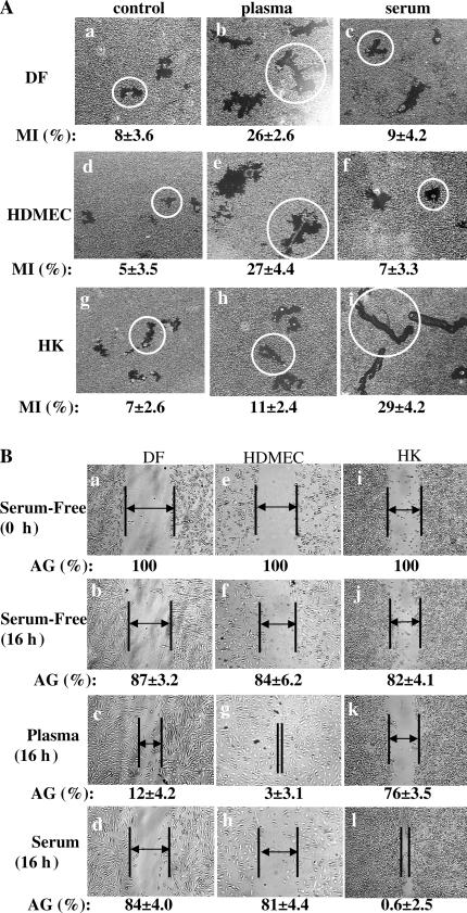
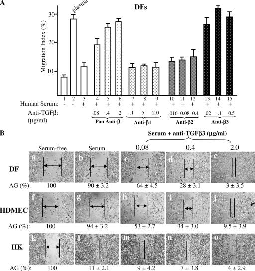
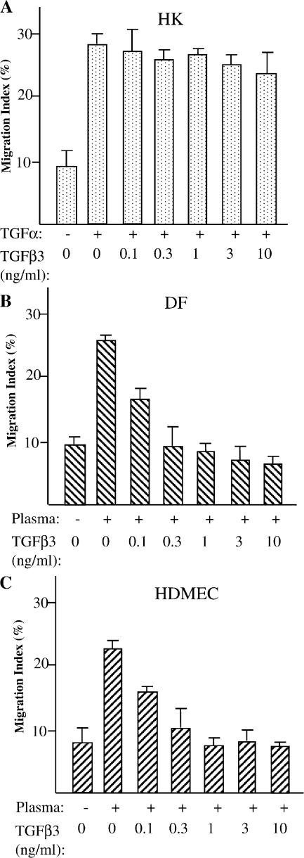
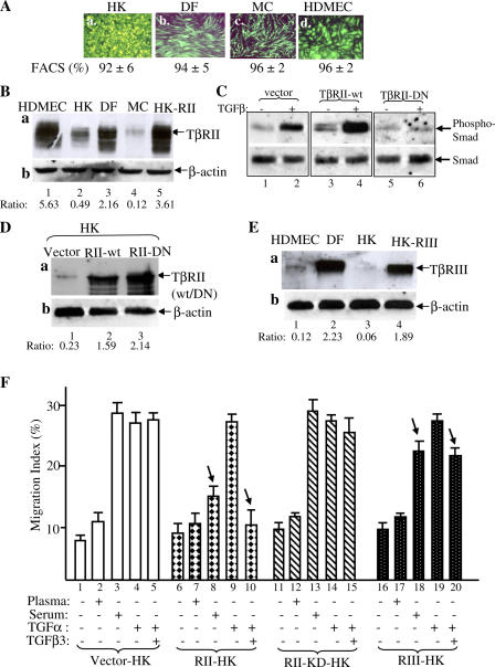
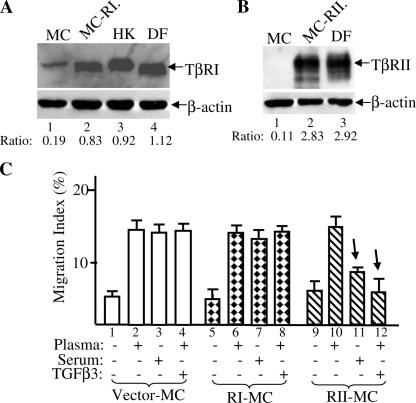
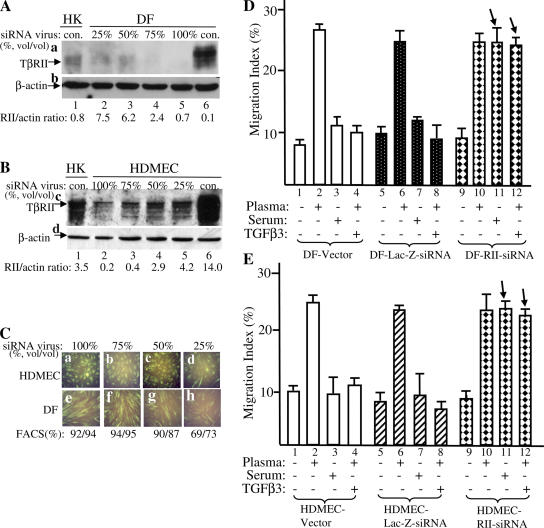
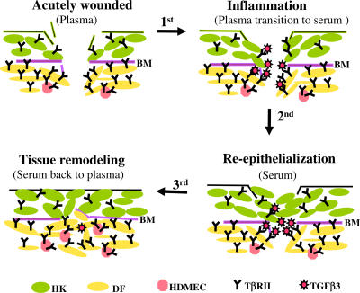
Cell migration is a rate-limiting event in skin wound healing. In unwounded skin, cells are nourished by plasma. When skin is wounded, resident cells encounter serum for the first time. As the wound heals, the cells experience a transition of serum back to plasma. In this study, we report that human serum selectively promotes epidermal cell migration and halts dermal cell migration. In contrast, human plasma promotes dermal but not epidermal cell migration. The on-and-off switch is operated by transforming growth factor (TGF) beta3 levels, which are undetectable in plasma and high in serum, and by TGFbeta receptor (TbetaR) type II levels, which are low in epidermal cells and high in dermal cells. Depletion of TGFbeta3 from serum converts serum to a plasmalike reagent. The addition of TGFbeta3 to plasma converts it to a serumlike reagent. Down-regulation of TbetaRII in dermal cells or up-regulation of TbetaRII in epidermal cells reverses their migratory responses to serum and plasma, respectively. Therefore, the naturally occurring plasma-->serum-->plasma transition during wound healing orchestrates the orderly migration of dermal and epidermal cells.


Figures

Figure 1.
Figure 1.
Plasma and serum oppositely affect dermal and epidermal cell migration. HKs, DFs, and HDMECs were serum starved overnight and subjected to colloidal gold migration assays (A) and in vitro wound-healing assays (B). (A) Representative images of the colloidal gold migration assays under the following three conditions: (1) on a collagen matrix in the absence of any soluble GFs (a, d, and g); (2) on a collagen matrix in the presence of an optimal concentration (10% vol/vol) of human plasma (b, e, and h); and (3) on collagen matrix in the presence of an optimal concentration (10% vol/vol) of human serum (c, f, and l). The computer-assisted quantitative analyses of the migration tracks are shown as migration indices (MI). These experiments were repeated five times, and closely similar results were obtained. Average size migration tracks are marked with circles. (B) In vitro wound-healing assay was performed under the same conditions. The wound closures were photographed, and the average gaps (AGs) were quantitated as described previously (Li et al., 2004b). AGs are indicated by lines and arrows; some gaps were too close to insert an arrow. Similar results were reproduced in three independent experiments.
Figure 2.
Figure 2.
TGFβ3 but not TGFβ1 or TGFβ2 in human serum selectively blocks dermal cell migration. DFs, HDMECs, and HKs were serum starved overnight and subjected to migration assays on collagen in the absence or presence of plasma, serum, or serum with added increasing amounts of anti-TGFβ neutralizing antibodies. The four antibodies include a pan-antibody against all three TGFβ isoforms and three other antibodies specifically against the individual TGFβ1, TGFβ2, or TGFβ3. The amounts of these antibodies were predetermined to neutralize the same amounts of their corresponding TGFβ isoforms. (A) Anti-TGFβ3 antibody released the inhibition of serum in DF migration, whereas anti-TGFβ1 and anti-TGFβ2 antibodies did not. This experiment was repeated four times. Error bars represent SEM. (B) The addition of anti-TGFβ3 antibody to serum converted to a promotility agent for both DFs and HDMECs. The cells were subjected to in vitro wound-healing assays under either serum-free, serum, or serum with added anti-TGFβ3 neutralizing antibodies. After 16 h, the wound closures were photographed, and the AGs were quantitated as described previously (Li et al., 2004b). AGs are indicated by lines and arrows; some gaps were too close to insert an arrow. The results shown here were reproducible in two independent experiments.
Figure 3.
Figure 3.
TGFβ3 selectively inhibits dermal, but not epidermal, cell migration. DFs, HDMECs, and HKs were serum starved overnight and subjected to colloidal gold migration assays on collagen. The indicated concentrations of TGFβ3 were added to either TGFα- (stimulus of HK migration to replace serum that already contains TGFβ3) or plasma (stimulus of DF and HDMEC migration)-containing media. MIs of the migration are shown. The differences in MIs in HKs were statistically insignificant (P < 0.001). The differences in MIs in DFs and HDMECs were statistically significant (P < 0.005). These experiments were repeated four times, and reproducible results were obtained. Error bars represent SEM.
Figure 4.
Figure 4.
In vitro and in vivo human skin profiles of TβRs (I, II, and III) and their transmission of TGFβ-stimulated Smad2/3 activation. (A) Equal amounts of cell lysates of serum-starved HKs, MCs, DFs, and HDMECs were resolved in three SDS gels and subjected to Western blot analyses with antibodies against TβRI/ALK5 (a), TβRII (c), or TβRIII/betaglycan (e). Each lower part of the three membranes was blotted with an anti–β-actin antibody as a control (b, d, and f). The expression was quantitated by a densitometry scan, giving rise to a ratio of TβR band intensity over its corresponding β-actin band intensity. (B) Normal human skin sections were subjected to indirect immunofluorescence staining with the antibodies against TβRI (a), TβRII (f), TβRIII (k), DAPI (b, g, and l), rhodamine-conjugated phalloidin (c, h, and m), or corresponding IgG controls (see Materials and methods). The images show skin tissue distribution of the TβRs (a, f, and k), nuclei (b, g, and l), F-actin (c, h, and m), and all three stains merged (d, i, and n). A section of the anti-TβRI antibody staining (a, dotted box) was enlarged in the inset to visualize cellular levels of staining. Bar, 20 μm. (C) The cells were either untreated or treated with the indicated concentrations of TGFβ3 for 15 min, and equalized cell lysates (40 μg/lane) were subjected to Western blot analyses with either antiphospho-Smad2/3 (a, c, and e) or anti-Smad2/3 (b, d, and f) antibodies. (D) The cells were either untreated or treated with 2.5 ng/ml TGFβ3 for the indicated periods of time. Equalized cell lysates were subjected to Western blot analyses with either antiphospho-Smad2/3 (a, c, and e) or anti-Smad2/3 (b, d, and f) antibodies.
Figure 5.
Figure 5.
Up-regulation of TβRII converts epidermal HK motility to become TGFβ3 sensitive just like dermal cells. (A) Lentiviral infection offered >90% gene transduction efficiency in DFs, MCs, HDMECs, and HKs. 48 h after infection with pRRLsin-CMV-EGFP, the plates were analyzed under fluorescence microscopy and photographed. The cells were suspended by trypsin and subjected to FACS analyses for measurement of the gene transduction efficiency (percentage of green cells/total cells). (B) Elevated expression of TβRII in HKs after pRRLsin-TβRII infection (lane 5) in comparison with the endogenous TβRII expression in HKs (lane 2), DFs (lane 3), and HDMECs (lane 1). Equal amounts of cell extracts of the indicated cell types were blotted with anti-TβRII antibody (a) or anti–β-actin antibody (b). (C) TβRII-KD blocks TGFβ3-stimulated Smad2/3 phosphorylation in HKs. The HKs, infected with vector, wild-type TβRII, or TβRII-KD, were untreated or treated with 2.5 ng/ml TGFβ3 for 45 min and analyzed for the phosphorylation of Smad2/3 similar to the assays shown in Fig. 4 (C and D). (D) Elevated expression of TβRII-KD in HKs after pRRLsin-CMV–TβRII-KD infection (lane 3) in comparison with elevated wild-type TβRII expression (lane 2) and the endogenous TβRII expression in HKs (lane 1). (E) Elevated expression of TβRIII in HKs (lane 4 vs. lane 3) after pRRLsin-CMV-TβRIII infection in comparison with endogenous TβRIII expression in DFs (lane 2) and HDMECs (lane 1). (F) The aforementioned TβR-engineered HKs were tested for their migratory responses to plasma, serum, or TGFα with or without 0.5 ng/ml TGFβ3 in colloidal gold migration assays. MIs are shown. This experiment was repeated three times. Error bars represent SEM.
Figure 6.
Figure 6.
Up-regulation of TβRII, but not TβRI, causes epidermal MC motility to become TGFβ3 sensitive. (A) Elevated expression of TβRI in MCs (lane 2 vs. lane 1) after pRRLsin-CMV-TβRI infection in comparison with endogenous TβRI expression in HKs (lane 3) and DFs (lane 4). (B) Elevated expression of TβRII in MCs (lane 2 vs. lane 1) after pRRLsin-CMV-TβRII infection in comparison with endogenous TβRII expression in DFs (lane 3). (C) Migration of MCs with vector control (bars 1–4), overexpressing TβRI (bars 5–8), or overexpressing TβRII (bars 9–12) in the presence of plasma, serum, or plasma plus 0.5 ng/ml TGFβ3. Error bars represent SEM.
Figure 7.
Figure 7.
Down-regulation of TβRII causes dermal DF and HDMEC motility to become TGFβ3 insensitive just like the epidermal cells. DFs and HDMECs were infected with various dilutions (vol/vol) of the lentivirus (FG-12) carrying an siRNA against TβRII. After 48 h, the cells were first analyzed for down-regulation of the endogenous TβRII before being subjected to migration assays. (A) 50% dilution of the original virus stock decreases TβRII expression in DFs (a, lane 6) to the level similar to HKs (a, lane 3 vs. lane 1). (B) The same dilution of the virus stock decreases the TβRII expression in HDMECs (c, lane 6) to the similar level in HKs (c, lane 3 vs. lane 1). (C) Coexpressed GFP and FACS analyses show no significant reduction in gene transduction efficiency in HDMECs (a–c) or DFs (e–g) unless the virus stock is diluted to 25% or further (d and h). (D and E) DF and HDMEC cells 48 h after infection with 50% of the original virus stocks of vector alone, lac-Z–siRNA, and TβRII-siRNA were subjected to colloidal gold migration assays in the absence or presence of plasma, serum, and plasma plus TGFβ3. MIs are shown. These migration experiments were repeated three times. Error bars represent SEM.
Figure 8.
Figure 8.
A schematic representation of how the plasma→serum→plasma transitions coordinate the orderly skin cell migration during wound healing. Three major types of skin cells—HKs, DFs, and HDMECs—are shown here, as indicated by different colors. The dermal cells express higher levels of TβRII (symbolized by Y) than the epidermal cells. Therefore, the dermal cells are sensitive to the anti-promotility effect of TGFβ3 (red stars), whose concentration increases after the transition from plasma to serum in the wound bed. Contributions by other cell types and matrix components to skin wound healing are omitted for the sake of simplicity but not reality. The relative numbers and proportions of the various types of cells do not quantitatively reflect those in real human skin. BM, basement membrane.

References

    1. Albrecht-Buehler, G. 1977. The phagokinetic tracks of 3T3 cells. Cell. 11:395–404. - PubMed
    1. Ashcroft, G.S., X. Yang, A.B. Glick, M. Weinsteil, J.L. Letterio, D.E. Mizel, M. Anzano, T. Greenwell-Wild, S.M. Wahl, C. Deng, and A.B. Roberts. 1999. Mice lacking Smad3 show accelerated wound healing and an impaired local inflammatory response. Nat. Cell Biol. 1:260–266. - PubMed
    1. Assoian, R.K., A. Komoriya, C.A. Meyers, D.M. Miller, and M.B. Sporn. 1983. Transforming growth factor-beta in human platelets. Identification of a major storage site, purification, and characterization. J. Biol. Chem. 258:7155–7160. - PubMed
    1. Assoian, R.K., G.R. Grotendorst, D.M. Miller, and M.B. Sporn. 1984. Cellular transformation by coordinated action of three peptide growth factors from human platelets. Nature. 309:804–806. - PubMed
    1. Assoian, R.K., B.E. Fleurdelys, H.C. Stevenson, P.J. Miller, D.K. Madtes, E.W. Raines, R. Ross, and M.B. Sporn. 1987. Expression and secretion of type beta transforming growth factor by activated human macrophages. Proc. Natl. Acad. Sci. USA. 84:6020–6024. - PMC - PubMed

Publication types

MeSH terms