. 2006 Mar 27;172(7):1023-34.
doi: 10.1083/jcb.200507116. Epub 2006 Mar 20.

GPI-anchored proteins are directly targeted to the apical surface in fully polarized MDCK cells

Affiliations

GPI-anchored proteins are directly targeted to the apical surface in fully polarized MDCK cells

Simona Paladino et al. J Cell Biol. .

Abstract

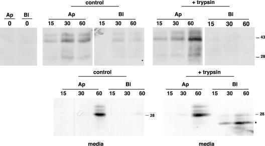
The polarity of epithelial cells is dependent on their ability to target proteins and lipids in a directional fashion. The trans-Golgi network, the endosomal compartment, and the plasma membrane act as sorting stations for proteins and lipids. The site of intracellular sorting and pathways used for the apical delivery of glycosylphosphatidylinositol (GPI)-anchored proteins (GPI-APs) are largely unclear. Using biochemical assays and confocal and video microscopy in living cells, we show that newly synthesized GPI-APs are directly delivered to the apical surface of fully polarized Madin-Darby canine kidney cells. Impairment of basolateral membrane fusion by treatment with tannic acid does not affect the direct apical delivery of GPI-APs, but it does affect the organization of tight junctions and the integrity of the monolayer. Our data clearly demonstrate that GPI-APs are directly sorted to the apical surface without passing through the basolateral membrane. They also reinforce the hypothesis that apical sorting of GPI-APs occurs intracellularly before arrival at the plasma membrane.


Figures

Figure 1.
Figure 1.
Newly synthesized GPI-APs are directly targeted to the apical surface. MDCK cells stably expressing GFP-GPI or PLAP grown on filters for 4 (A) or 1.5 d (B) were pulsed for 15 min with [35S]methionine and chased for the indicated times. At the end of each chase time, cells were labeled with LC-biotin and added to the apical (Ap) or basolateral (Bl) surface, respectively. Cell lysates were immunoprecipitated with specific antibodies. The biotinylated proteins were recovered from the immunoprecipitates by reprecipitation with streptavidin beads. After running on SDS-PAGE, samples were analyzed by phosphorimager. One tenth of immunoprecipitates (totals) was kept before streptavidin precipitation and shown in the bottom panels. The monomer form of GFP-GPI (28 kD) and the slower mobility band (43 kD), a partially denaturated GFP dimer that was previously described (Inouye and Tsuji, 1994; Paladino et al., 2004), are indicated. Fluorograms of four different experiments were quantified, and the results were expressed as a percentage of the amount at the time of maximal surface expression. For GFP-GPI, the quantification is referred to as the upper and lower bands together. (C) MDCK cells stably expressing GFP-GPI grown on filters for 1, 2.5, and 4 d were pulsed for 15 min with [35S]methionine, chased for 30, 60, and 90 min, and processed as in A and B. Results from four different experiments were plotted as percent apical and basolateral surface expression. Error bars represent SD.
Figure 2.
Figure 2.
GFP-GPI is directly delivered from the Golgi to the apical surface. MDCK cells stably expressing GFP-GPI grown on filter for 4 d were incubated with trypsin to eliminate all proteins at the surface and were subjected to a temperature block to accumulate proteins in the TGN (see Materials and methods). Cells were then warmed at 37°C for the indicated times in culture medium containing cycloheximide. After each time point, cells were fixed and treated for confocal microscopy where serial confocal sections were collected from the top to the bottom of the cell monolayer (A). The dashed lines show the positions from where the xz sections were taken. Mean fluorescence intensities at the Golgi, apical, and basolateral domains were measured at the different chase times and expressed as percentages of total fluorescence (A, bottom; see Materials and methods). Alternatively, cells were biotinylated from the apical (Ap) or basolateral (Bl) side. After lysis, GFP-GPI was immunoprecipitated with an antibody against GFP and revealed by using HRP-streptavidin (B). Gels from three different experiments were quantified, and the results were expressed as percentages of the amount at the time of maximal surface expression. The amount of protein still present at the apical or basolateral surface at time = 0 was subtracted from all chase times. Error bars represent SD. Bars, 10 μm.
Figure 3.
Figure 3.
GFP-GPI still reaches the apical surface in the presence of trypsin in the basolateral medium. MDCK cells stably expressing GFP-GPI grown on filter for 4 d were incubated with trypsin, subjected to a temperature block in the TGN (see Materials and methods), and chased at 37°C for the indicated times (in minutes) in the presence or absence of 25 μg/ml trypsin in the basolateral media. After each chase time, cells were selectively biotinylated from the apical (Ap) or basolateral (Bl) surface. After immunoprecipitation with an antibody against GFP, samples were run on SDS-PAGE and revealed using HRP-streptavidin. Apical and basolateral media for control and treated cells were recovered at all chase times, proteins were TCA precipitated, run on SDS-PAGE, and revealed by Western blotting using GFP antibody. The asterisk indicates the trypsin partially digested form of GFP-GPI.
Figure 4.
Figure 4.
Tannic acid does not affect the apical targeting of GFP-GPI. MDCK cells stably expressing GFP-GPI grown on filter were incubated with trypsin and subjected to a temperature block in the TGN (see Materials and methods). Tannic acid was added to the basolateral medium 30 min before release from the 20°C block. Cells were then warmed at 37°C for the indicated times in the absence of tannic acid in culture medium containing cycloheximide. After each time point, cells were fixed and quenched. Serial confocal sections were collected from the top to the bottom of cell monolayers (A). The dashed lines show the positions from where the xz sections were taken. Mean fluorescence intensities at the Golgi, apical (Ap), and basolateral (Bl) domains were measured as in Fig. 2. Alternatively, surface proteins were biotinylated from the apical or basolateral side, immunoprecipitated with GFP antibody, and revealed by using HRP-streptavidin (B). Results from three different experiments were plotted as the percentage of the amount at the time of maximal surface expression. The value at the apical or basolateral surface at time = 0 was subtracted from all chase times. Error bars represent SD. Bars, 10 μm.
Figure 5.
Figure 5.
Long tannic acid treatment leads to the depolarization of GFP-GPI. MDCK cells stably expressing GFP-GPI grown on filter were incubated with trypsin and subjected to a temperature block in the TGN. Tannic acid was added to the basolateral medium 10 min before release from the 20°C block. Cells were warmed at 37°C for the indicated times in culture medium containing cycloheximide in the presence of tannic acid. After each time point, cells were fixed, quenched, and analyzed by confocal microscopy. Mean fluorescence intensities at the Golgi, apical (Ap), and basolateral (Bl) domains were measured as in Fig. 2. Alternatively, surface proteins were biotinylated from the apical or the basolateral side, immunoprecipitated with GFP antibody, and revealed by using HRP-streptavidin (B). Results from three different experiments were plotted as percentages of the amount at the time of maximal surface expression. The value at the apical or basolateral surface at time = 0 was subtracted from all chase times. Error bars represent SD. Bars, 10 μm.
Figure 6.
Figure 6.
Long tannic acid treatment alters the tight junctions and affects the integrity of the monolayer. Tannic acid was added to MDCK cells grown on filters for 30 or 60 min at 37°C. (A) Cells were then fixed and stained with specific antibodies against PATJ (green) and Na,K-ATPase (red) in permeabilized conditions. Serial confocal sections were collected from the top to the bottom of cell monolayers. (B) After culturing MDCK cells on filter for 4 d, media were replaced, and transepithelial resistance (TER) was measured periodically over 90 min in the absence (control) or presence of tannic acid added to the basolateral side. (C) [14C]inulin was added to the apical medium of MDCK cells grown on filter, whereas tannic acid was added (or not; control) to the basolateral medium for the indicated times at 37°C. The permeability of the monolayers was subsequently assessed by quantitating the percentage of apical [14C]inulin found in the basal well. The data are means ± SD (error bars) of three experiments. Bar, 10 μm.
Figure 7.
Figure 7.
GFP-GPI is directly delivered to the apical surface in living MDCK cells both in the presence and absence of tannic acid. MDCK cells stably expressing GFP-GPI were treated as in Figs. 2 and 4. After the temperature block, cells were shifted at 37°C on the microscope and imaged live every 3 min for 60 min (A) or every 1 min for 15 min (B; also see Videos 1–8, available at http://www.jcb.org/cgi/content/full/jcb.200507116/DC1).Some frames of the time lapses three-dimensionally reconstructed with different orientations (A [top] and B, view from the apical side; A [bottom], view from the lateral side) are shown. Bars, 10 μm.

Comment in

References

    1. Ang, A.L., T. Taguchi, S. Francis, H. Folsch, L.J. Murrells, M. Pypaert, G. Warren, and I. Mellman. 2004. Recycling endosomes can serve as intermediates during transport from the Golgi to the plasma membrane of MDCK cells. J. Cell Biol. 167:531–543. - PMC - PubMed
    1. Apodaca, G., and K.E. Mostov. 1993. Transcytosis of placental alkaline phosphatase-polymeric immunoglobulin receptor fusion proteins is regulated by mutations of Ser664. J. Biol. Chem. 268:23712–23719. - PubMed
    1. Apodaca, G., M. Bomsel, R. Lindstedt, J. Engel, D. Frank, K.E. Mostov, and J. Wiener-Kronish. 1995. Characterization of Pseudomonas aeruginosa-induced MDCK cell injury: glycosylation-defective host cells are resistant to bacterial killing. Infect. Immun. 63:1541–1551. - PMC - PubMed
    1. Arreaza, G., and D.A. Brown. 1995. Sorting and intracellular trafficking of a glycosylphosphatidylinositol-anchored protein and two hybrid transmembrane proteins with the same ectodomain in Madin-Darby canine kidney epithelial cells. J. Biol. Chem. 270:23641–23647. - PubMed
    1. Bonifacino, J.S., and L.M. Traub. 2003. Signals for sorting of transmembrane proteins to endosomes and lysosomes. Annu. Rev. Biochem. 72:395–447. - PubMed

Publication types

MeSH terms