

Serpin squamous cell carcinoma antigen inhibits UV-induced apoptosis via suppression of c-JUN NH2-terminal kinase
- PMID: 16549498
- PMCID: PMC2063756
- DOI: 10.1083/jcb.200508064
Serpin squamous cell carcinoma antigen inhibits UV-induced apoptosis via suppression of c-JUN NH2-terminal kinase
Abstract
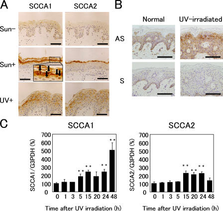
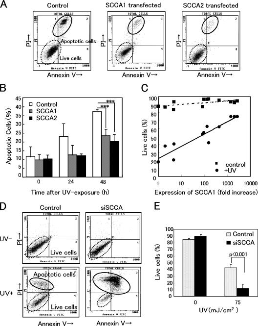
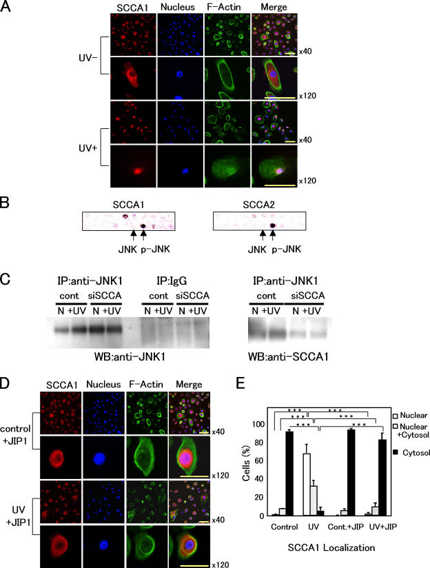
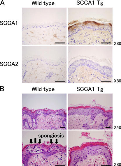
Protection from ultraviolet (UV) irradiation is a fundamental issue for living organisms. Although melanin's critical role in the protection of basal keratinocytes is well understood, other factors remain essentially unknown. We demonstrate that up-regulation of squamous cell carcinoma antigen-1 (SCCA1) suppresses c-Jun NH2-terminal kinase-1 (JNK1) and thus blocks UV-induced keratinocyte apoptosis. We found that serpin SCCA1 is markedly elevated in the top layers of sun-exposed or UV-irradiated epidermis. UV-induced apoptosis was significantly decreased when SCCA was overexpressed in 3T3/J2 cells. It was significantly increased when SCCA was down-regulated with small interfering RNA in HaCaT keratinocytes. A search for SCCA-interacting molecules showed specific binding with phosphorylated JNK. Interestingly, SCCA1 specifically suppressed the kinase activity of JNK1. Upon exposure of keratinocytes to UV, SCCA1 was bound to JNK1 and transferred to the nucleus. Involucrin promoter-driven SCCA1 transgenic mice showed remarkable resistance against UV irradiation. These findings reveal an unexpected serpin function and define a novel UV protection mechanism in human skin.
Figures

Similar articles
-
c-JUN N-terminal kinase-1 (JNK1) but not JNK2 or JNK3 is involved in UV signal transduction in human epidermis.J Dermatol Sci. 2006 Sep;43(3):171-9. doi: 10.1016/j.jdermsci.2006.05.008. Epub 2006 Jul 5. J Dermatol Sci. 2006. PMID: 16824735
-
Crystal structure of SCCA1 and insight about the interaction with JNK1.Biochem Biophys Res Commun. 2009 Feb 27;380(1):143-7. doi: 10.1016/j.bbrc.2009.01.057. Epub 2009 Jan 21. Biochem Biophys Res Commun. 2009. PMID: 19166818
-
WOX1 is essential for UVB irradiation-induced apoptosis and down-regulated via translational blockade in UVB-induced cutaneous squamous cell carcinoma in vivo.Clin Cancer Res. 2005 Aug 15;11(16):5769-77. doi: 10.1158/1078-0432.CCR-04-2274. Clin Cancer Res. 2005. PMID: 16115915
-
Expression and function of squamous cell carcinoma antigen.Anticancer Res. 1996 Jul-Aug;16(4B):2149-53. Anticancer Res. 1996. PMID: 8694535 Review.
-
SERPINB3, apoptosis and autoimmunity.Autoimmun Rev. 2009 Dec;9(2):108-12. doi: 10.1016/j.autrev.2009.03.011. Epub 2009 Mar 27. Autoimmun Rev. 2009. PMID: 19332150 Review.
Cited by
-
Epitope-Specific Anti-SerpinB3 Antibodies for SerpinB3 Recognition and Biological Activity Inhibition.Biomolecules. 2023 Apr 25;13(5):739. doi: 10.3390/biom13050739. Biomolecules. 2023. PMID: 37238609 Free PMC article.
-
Heparin enhances serpin inhibition of the cysteine protease cathepsin L.J Biol Chem. 2010 Feb 5;285(6):3722-3729. doi: 10.1074/jbc.M109.037358. Epub 2009 Dec 3. J Biol Chem. 2010. PMID: 19959474 Free PMC article.
-
Differential effects of periopathogens on host protease inhibitors SLPI, elafin, SCCA1, and SCCA2.J Oral Microbiol. 2010 May 4;2. doi: 10.3402/jom.v2i0.5070. J Oral Microbiol. 2010. PMID: 21523231 Free PMC article.
-
SCCA1/SERPINB3 promotes oncogenesis and epithelial-mesenchymal transition via the unfolded protein response and IL6 signaling.Cancer Res. 2014 Nov 1;74(21):6318-29. doi: 10.1158/0008-5472.CAN-14-0798. Epub 2014 Sep 11. Cancer Res. 2014. PMID: 25213322 Free PMC article.
-
Combined detection of Twist1, Snail1 and squamous cell carcinoma antigen for the prognostic evaluation of invasion and metastasis in cervical squamous cell carcinoma.Int J Clin Oncol. 2018 Apr;23(2):321-328. doi: 10.1007/s10147-017-1210-2. Epub 2017 Nov 3. Int J Clin Oncol. 2018. PMID: 29101499
References
-
- Barr, R.K., T.S. Kendrick, and M.A. Bogoyevitch. 2002. Identification of the critical features of a small peptide inhibitor of JNK activity. J. Biol. Chem. 277:10987–10997. - PubMed
-
- Bonny, C., A. Oberson, S. Negri, C. Sauser, and D.F. Schorderet. 2001. Cell-permeable peptide inhibitors of JNK: novel blockers of beta-cell death. Diabetes. 50:77–82. - PubMed
-
- Butterfield, L., B. Storey, L. Maas, and L.E. Heasley. 1997. c-Jun NH2-terminal kinase regulation of the apoptotic response of small cell lung cancer cells to ultraviolet radiation. J. Biol. Chem. 272:10110–10116. - PubMed
MeSH terms
Substances
LinkOut - more resources
Full Text Sources
Other Literature Sources
Molecular Biology Databases
Research Materials
Miscellaneous
