In vivo analysis of the role of aberrant histone deacetylase recruitment and RAR alpha blockade in the pathogenesis of acute promyelocytic leukemia
- PMID: 16549595
- PMCID: PMC2118271
- DOI: 10.1084/jem.20050616
In vivo analysis of the role of aberrant histone deacetylase recruitment and RAR alpha blockade in the pathogenesis of acute promyelocytic leukemia
Abstract
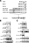
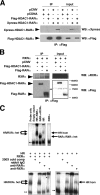
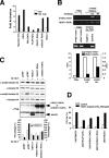
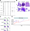
The promyelocytic leukemia-retinoic acid receptor alpha (PML-RARalpha) protein of acute promyelocytic leukemia (APL) is oncogenic in vivo. It has been hypothesized that the ability of PML-RARalpha to inhibit RARalpha function through PML-dependent aberrant recruitment of histone deacetylases (HDACs) and chromatin remodeling is the key initiating event for leukemogenesis. To elucidate the role of HDAC in this process, we have generated HDAC1-RARalpha fusion proteins and tested their activity and oncogenicity in vitro and in vivo in transgenic mice (TM). In parallel, we studied the in vivo leukemogenic potential of dominant negative (DN) and truncated RARalpha mutants, as well as that of PML-RARalpha mutants that are insensitive to retinoic acid. Surprisingly, although HDAC1-RARalpha did act as a bona fide DN RARalpha mutant in cellular in vitro and in cell culture, this fusion protein, as well as other DN RARalpha mutants, did not cause a block in myeloid differentiation in vivo in TM and were not leukemogenic. Comparative analysis of these TM and of TM/PML(-/-) and p53(-/-) compound mutants lends support to a model by which the RARalpha and PML blockade is necessary, but not sufficient, for leukemogenesis and the PML domain of the fusion protein provides unique functions that are required for leukemia initiation.
Figures


Similar articles
-
Fusion proteins of the retinoic acid receptor-alpha recruit histone deacetylase in promyelocytic leukaemia.Nature. 1998 Feb 19;391(6669):815-8. doi: 10.1038/35901. Nature. 1998. PMID: 9486655
-
The self-association coiled-coil domain of PML is sufficient for the oncogenic conversion of the retinoic acid receptor (RAR) alpha.Leukemia. 2011 May;25(5):814-20. doi: 10.1038/leu.2011.18. Epub 2011 Feb 18. Leukemia. 2011. PMID: 21331069
-
Acute leukemia with promyelocytic features in PML/RARalpha transgenic mice.Proc Natl Acad Sci U S A. 1997 May 13;94(10):5302-7. doi: 10.1073/pnas.94.10.5302. Proc Natl Acad Sci U S A. 1997. PMID: 9144232 Free PMC article.
-
Transcriptional regulation in acute promyelocytic leukemia.Oncogene. 2001 Oct 29;20(49):7204-15. doi: 10.1038/sj.onc.1204853. Oncogene. 2001. PMID: 11704848 Review.
-
Understanding the molecular pathogenesis of acute promyelocytic leukemia.Best Pract Res Clin Haematol. 2014 Mar;27(1):3-9. doi: 10.1016/j.beha.2014.04.006. Epub 2014 Apr 13. Best Pract Res Clin Haematol. 2014. PMID: 24907012 Review.
Cited by
-
All-trans retinoic acid is capable of inducing folate receptor β expression in KG-1 cells.Tumour Biol. 2010 Dec;31(6):589-95. doi: 10.1007/s13277-010-0074-0. Epub 2010 Jul 15. Tumour Biol. 2010. PMID: 20632143
-
Therapeutic Strategies Targeting Cancer Stem Cells and Their Microenvironment.Front Oncol. 2019 Oct 24;9:1104. doi: 10.3389/fonc.2019.01104. eCollection 2019. Front Oncol. 2019. PMID: 31709180 Free PMC article. Review.
-
Targeted Cancer Stem Cell Therapeutics: An Update.Curr Top Med Chem. 2025;25(8):842-870. doi: 10.2174/0115680266275014240110071351. Curr Top Med Chem. 2025. PMID: 38288804 Review.
-
The cell biology of disease: Acute promyelocytic leukemia, arsenic, and PML bodies.J Cell Biol. 2012 Jul 9;198(1):11-21. doi: 10.1083/jcb.201112044. J Cell Biol. 2012. PMID: 22778276 Free PMC article. Review.
-
The SUMO E3-ligase PIAS1 regulates the tumor suppressor PML and its oncogenic counterpart PML-RARA.Cancer Res. 2012 May 1;72(9):2275-84. doi: 10.1158/0008-5472.CAN-11-3159. Epub 2012 Mar 9. Cancer Res. 2012. PMID: 22406621 Free PMC article.
References
-
- Melnick, A., and J.D. Licht. 1999. Deconstructing a disease: RARα, its fusion partners, and their roles in the pathogenesis of acute promyelocytic leukemia. Blood. 93:3167–3215. - PubMed
-
- Zelent, A., F. Guidez, A. Melnick, S. Waxman, and J.D. Licht. 2001. Translocations of the RARα gene in acute promyelocytic leukemia. Oncogene. 20:7186–7203. - PubMed
-
- He, L.Z., F. Guidez, C. Tribioli, D. Peruzzi, M. Ruthardt, A. Zelent, and P.P. Pandolfi. 1998. Distinct interactions of PML-RARα and PLZF-RARα with co-repressors determine differential responses to RA in APL. Nat. Genet. 18:126–135. - PubMed
-
- Cheng, G.X., X.H. Zhu, X.Q. Men, L. Wang, Q.H. Huang, X.L. Jin, S.M. Xiong, J. Zhu, W.M. Guo, J.Q. Chen, et al. 1999. Distinct leukemia phenotypes in transgenic mice and different corepressor interactions generated by promyelocytic leukemia variant fusion genes PLZF-RARα and NPM-RARα. Proc. Natl. Acad. Sci. USA. 96:6318–6323. - PMC - PubMed
Publication types
MeSH terms
Substances
Grants and funding
LinkOut - more resources
Full Text Sources
Molecular Biology Databases
Research Materials
Miscellaneous

