. 2006 Mar 28;103(13):5161-6.
doi: 10.1073/pnas.0600948103. Epub 2006 Mar 20.

Early-onset behavioral and synaptic deficits in a mouse model of Alzheimer's disease

Affiliations

Early-onset behavioral and synaptic deficits in a mouse model of Alzheimer's disease

J Steven Jacobsen et al. Proc Natl Acad Sci U S A. .

Abstract

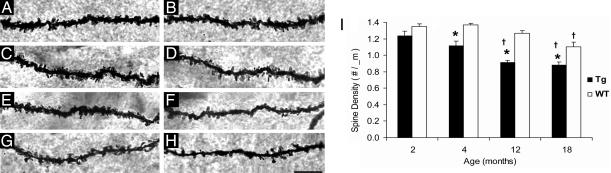
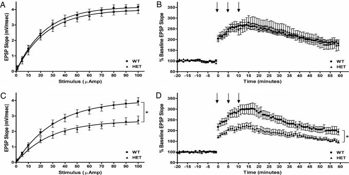
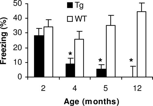
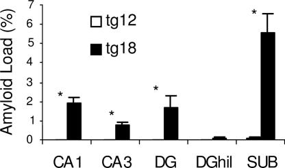
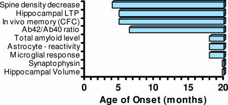
Alzheimer's disease (AD) is a progressive neurodegenerative disorder for which numerous mouse models have been generated. In both AD patients and mouse models, there is increasing evidence that neuronal dysfunction occurs before the accumulation of beta-amyloid (Abeta)-containing plaques and neurodegeneration. Characterization of the timing and nature of preplaque dysfunction is important for understanding the progression of this disease and to identify pathways and molecular targets for therapeutic intervention. Hence, we have examined the progression of dysfunction at the morphological, functional, and behavioral levels in the Tg2576 mouse model of AD. Our data show that decreased dendritic spine density, impaired long-term potentiation (LTP), and behavioral deficits occurred months before plaque deposition, which was first detectable at 18 months of age. We detected a decrease in spine density in the outer molecular layer of the dentate gyrus (DG) beginning as early as 4 months of age. Furthermore, by 5 months, there was a decline in LTP in the DG after perforant path stimulation and impairment in contextual fear conditioning. Moreover, an increase in the Abeta42/Abeta40 ratio was first observed at these early ages. However, total amyloid levels did not significantly increase until approximately 18 months of age, at which time significant increases in reactive astrocytes and microglia could be observed. Overall, these data show that the perforant path input from the entorhinal cortex to the DG is compromised both structurally and functionally, and this pathology is manifested in memory defects long before significant plaque deposition.


Conflict of interest statement

Conflict of interest statement: No conflicts declared.

Figures

Fig. 1.
Fig. 1.
Spine density in the DG. Spine segments are shown from 2-, 4-, 12-, and 18-month-old WT littermates (A, C, E, and G, respectively) and Tg mice (B, D, F, and H, respectively). Segments are from the molecular layer of the DG (middle and outer molecular layers combined). (Scale bar, 10 μm.) (I) Spine deficits were detected in 4-, 12-, and 18-month-old Tg mice compared with WT littermates but not in 2-month-old Tg mice (∗, P < 0.05). Also shown is a reduced spine density detected over time in Tg mice and WT littermates (†, P < 0.05; see Results for specific comparisons).
Fig. 2.
Fig. 2.
Basal synaptic transmission and LTP. The DG is synaptically impaired in 4- but not 2-month-old Tg mouse hippocampal slices. (A) I–O curves were derived from dendritic recordings of the DG after step-wise increases in perforant path stimulation. No difference was found between 2-month-old Tg and WT slices. (B) LTP was induced by multiple high-frequency stimulus trains. The level of potentiation was comparable between 2-month-old Tg and WT slices. (C) The I–O curves of 4- to 5-month-old slices were significantly impaired compared with age-matched WT controls. (D) Slices from 4- to 5-month-old Tg mice potentiated significantly less than those from age-matched WT controls. In B and D, arrows indicate application time points of conditioning stimulus trains. In C and D, statistical significance is denoted (∗, P < 0.01).
Fig. 3.
Fig. 3.
Age-associated impairment in CFC. Tg mice display intact contextual memory at 2 months of age. A significant impairment in hippocampal-dependent conditioning is observed with lower levels of freezing to context at 5-month-old Tg mice when compared with younger ages (∗, P < 0.05) with a trend toward an impairment in the 4-month-old group (†, P = 0.05). No differences were observed in freezing to context in the WT animals across all ages examined.
Fig. 4.
Fig. 4.
Tg brain Aβ42 levels. Quantitative analysis of Aβ42 and Aβ40 levels extracted from Tg mouse brain. Brain Aβ42 and Aβ40 was measured by using sandwich ELISA in mice from 2- to 8-month-old animals. Statistical analysis indicates that the percentage of Aβ42 rises significantly after 7 months relative to total Aβ (Aβ40 and Aβ42) levels (∗, P < 0.05).
Fig. 5.
Fig. 5.
Quantitative analysis of Aβ accumulation detected with 6E10. Caudal hippocampus is shown from 12- (on the left, open bars) and 18-month-old (on the right, closed bars) Tg mice. Significant Aβ accumulation was detected in 18-month-old mice in all subfields examined compared with minimal levels seen in 12-month-old mice. The subiculum contained significantly more Aβ compared with all other subfields (P < 0.05).
Fig. 6.
Fig. 6.
Temporal progression of morphological and functional deficits in Tg mice. The earliest onset of deficits in these mice occurs at ≈4 months of age and includes a decrease in hippocampal spine density, LTP, and in vivo memory as measured by CFC, and an increase in the ratio of Aβ42/Aβ40. Slower onset deficits include an increase in amyloid load, reactive astrocytes, and microglia. No changes in synaptophysin staining or hippocampal volume were observed at any age up to 18-month-old animals.

References

    1. Selkoe D. J. Ann. N.Y. Acad. Sci. 2000;924:17–25. - PubMed
    1. Hardy J., Selkoe D. J. Science. 2002;297:353–356. - PubMed
    1. Bloom G. S., Ren K., Glabe C. G. Biochim. Biophys. Acta. 2005;1739:116–124. - PubMed
    1. Ashe K. H. Learn. Mem. 2001;8:301–308. - PubMed
    1. Morgan D. Neurochem. Res. 2003;28:1029–1034. - PubMed