. 2006 Mar 28;103(13):5084-9.
doi: 10.1073/pnas.0600548103. Epub 2006 Mar 20.

Familial hypercatabolic hypoproteinemia caused by deficiency of the neonatal Fc receptor, FcRn, due to a mutant beta2-microglobulin gene

Affiliations

Familial hypercatabolic hypoproteinemia caused by deficiency of the neonatal Fc receptor, FcRn, due to a mutant beta2-microglobulin gene

Manzoor A Wani et al. Proc Natl Acad Sci U S A. .

Erratum in

Abstract

Two siblings, products of a consanguineous marriage, were markedly deficient in both albumin and IgG because of rapid degradation of these proteins, suggesting a lack of the neonatal Fc receptor, FcRn. FcRn is a heterodimeric receptor composed of a nonclassical MHC class I alpha-chain and beta(2)-microglobulin (beta(2)m) that binds two ligands, IgG and albumin, and extends the catabolic half-lives of both. Eight relatives of the siblings were moderately IgG-deficient. From sera archived for 35 years, we sequenced the two siblings' genes for the heterodimeric FcRn. We found that, although the alpha-chain gene sequences of the siblings were normal, the beta(2)m genes contained a single nucleotide transversion that would mutate a conserved alanine to proline at the midpoint of the signal sequence. Concentrations of soluble beta(2)m and HLA in the siblings' sera were <1% of normal. Transfection assays of beta(2)m-deficient cultured cells with beta(2)m cDNA indicated that the mutant beta(2)m supported <20% of normal expression of beta(2)m, MHC class I, and FcRn proteins. We concluded that a beta(2)m gene mutation underlies the hypercatabolism and reduced serum levels of albumin and IgG in the two siblings with familial hypercatabolic hypoproteinemia. This experiment of nature affirms our hypothesis that FcRn binds IgG and albumin, salvages both from a degradative fate, and maintains their physiologic concentrations.


Conflict of interest statement

Conflict of interest statement: No conflicts declared.

Figures

Fig. 1.
Fig. 1.
The β2m gene mutation in FHH patients. (a) Electropherograms comparing the nucleotide sequences of β2m exon 1 DNA amplified from serum samples of three normal individuals (N1, N2, and N3) and the two patients (S1 and S2). The emboldened base in the forward sequences identifies the single nucleotide mutation from G to C in the two FHH patients. The reverse strand sequence complements the forward sequence. (b) An ethidium-stained separation gel showing HinP1I (G/CGC) restriction enzyme digests of a 488-bp segment of exon 1 DNA sequence of the β2m gene PCR-amplified from relevant DNA samples. Marginal numbers indicate fragment sizes in bp. DNA from three normal sera and from cells of one normal individual (CD) show a wt restriction pattern with six restriction fragments (2, 13, 72, 79, 100, and 222 bp), whereas DNA from the patients show a diagnostically distinctive pattern of five restriction fragments (2, 13, 79,100, and 294 bp). (c) Restriction map of the PCR-amplified 488-bp segment. The vertical lines denote the HinP1I restriction sites; the numbers indicate the resulting fragment sizes. The red x marks the mutated (and thereby lost) HinP1I restriction site in the DNA of the two FHH patients, surrounding the point mutation (GCGC/CCGC) and causing the loss of the 72-bp fragment and the gain of the 294-bp fragment. (d) The 20-aa signal sequence (to arrow) of the β2m protein, with corresponding codons. The box indicates the single nucleotide transversion from G in the normal sequence (red) to C in the FHH patient sequence (blue). This transversion results in a single amino acid mutation from alanine to proline.
Fig. 2.
Fig. 2.
Overexpression of FHH mutant β2m fails to restore MHC I and β2m expression in a β2m-deficient cell line. A β2m-deficient cell line was transfected with a plasmid containing wt hβ2m cDNA (black), FHH mutant hβ2m cDNA (red), or plasmid alone (blue). Cells were stained with anti-β2m antibody (a), with anti-MHC I antibody (b), and with IgG isotype-matched control antibodies, followed by FITC-labeled goat anti-mouse secondary antibody (c and d). The results of one of the three representative flow cytometry experiments are expressed as histograms of fluorescence intensity (log scale) vs. the number of cells.
Fig. 3.
Fig. 3.
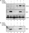
Mutant β2m fails to enhance FcRn expression. (a) Analysis of lysates. Nonionic detergent lysates of β2m-deficient cultured cells (FO-1) transfected with tabulated combinations of wt hβ2m cDNA, mut hβ2m cDNA, FcRn α-chain cDNA, and vectors alone (all lanes 1), were analyzed by SDS/PAGE and immunoblotting with rabbit anti-human FcRn antibody, rabbit anti-human β2m antibody, and anti-β-actin antibody as a protein-loading control. (b) Immunoadsorption of β2m. Lysates in OG buffer (pH 7.5) were immunoadsorbed with anti-hβ2m mAb (BBM.1). Eluted proteins were immunoblotted with rabbit anti-hβ2m antibody.

References

    1. Simister N. E., Mostov K. E. Nature. 1989;337:184–187. - PubMed
    1. Chaudhury C., Mehnaz S., Robinson J. M., Hayton W. L., Pearl D. K., Roopenian D. C., Anderson C. L. J. Exp. Med. 2003;197:315–322. - PMC - PubMed
    1. Ghetie V., Hubbard J. G., Kim J. K., Tsen M. F., Lee Y., Ward E. S. Eur. J. Immunol. 1996;26:690–696. - PubMed
    1. Junghans R. P., Anderson C. L. Proc. Natl. Acad. Sci. USA. 1996;93:5512–5516. - PMC - PubMed
    1. Israel E. J., Wilsker D. F., Hayes K. C., Schoenfeld D., Simister N. E. Immunology. 1996;89:573–578. - PMC - PubMed

Publication types

MeSH terms