. 2006 Mar 28;103(13):5060-5.
doi: 10.1073/pnas.0511167103. Epub 2006 Mar 20.

Galectin-3 regulates myofibroblast activation and hepatic fibrosis

Affiliations

Galectin-3 regulates myofibroblast activation and hepatic fibrosis

Neil C Henderson et al. Proc Natl Acad Sci U S A. .

Abstract

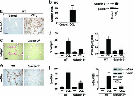
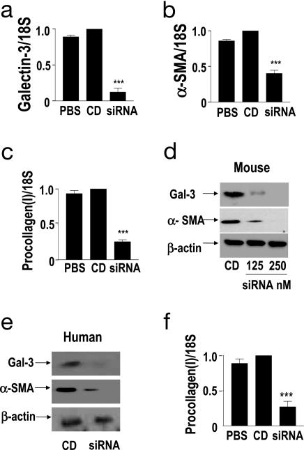
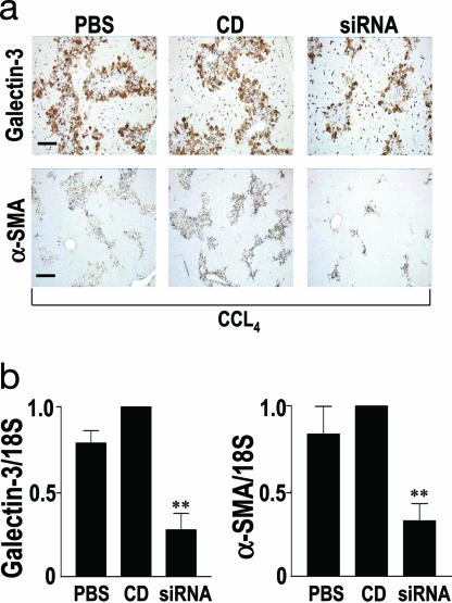
Central to fibrogenesis and the scarring of organs is the activation of fibroblasts into matrix-secreting myofibroblasts. We demonstrate that Galectin-3 expression is up-regulated in established human fibrotic liver disease and is temporally and spatially related to the induction and resolution of experimental hepatic fibrosis. Disruption of the Galectin-3 gene blocks myofibroblast activation and procollagen (I) expression in vitro and in vivo, markedly attenuating liver fibrosis. Addition of exogenous recombinant Galectin-3 in vitro reversed this abnormality. The reduction in hepatic fibrosis observed in the Galectin-3(-/-) mouse occurred despite equivalent liver injury and inflammation, and similar tissue expression of TGF-beta. TGF-beta failed to transactivate Galectin-3(-/-) hepatic stellate cells, in contrast with WT hepatic stellate cells; however, TGF-beta-stimulated Smad-2 and -3 activation was equivalent. These data suggest that Galectin-3 is required for TGF-beta mediated myofibroblast activation and matrix production. Finally, in vivo siRNA knockdown of Galectin-3 inhibited myofibroblast activation after hepatic injury and may therefore provide an alternative therapeutic approach to the prevention and treatment of liver fibrosis.


Conflict of interest statement

Conflict of interest statement: No conflicts declared.

Figures

Fig. 1.
Fig. 1.
Galectin-3 expression is up-regulated in human liver fibrosis. (a) Galectin-3 expression in normal human liver. (b) Galectin-3 in cirrhosis secondary to hepatitis C infection. (Scale bar: 400 μm.) Galectin-3 expression is temporally and spatially related to fibrosis in a reversible rat model of liver fibrosis. (c Upper) Collagen stained with PSR. (Scale bar: 100 μm.) (c Lower) Galectin-3 immunohistochemistry. (Scale bar: 200 μm.) (Left) Control (olive oil vehicle only). (Middle) Peak fibrosis in rat liver after 12 weeks of twice weekly i.p. CCL4. (Right) Resolution, 24 weeks after cessation of CCL4-induced liver injury.
Fig. 2.
Fig. 2.
Galectin-3 plays a critical role in organ fibrosis. Mice were treated with olive oil (control) or CCL4 i.p. twice weekly for 8 weeks (n = 6 mice in each group). (a) Galectin-3 expression in control (Left) and after chronic CCL4 treatment (Right) in WT mouse liver. (Scale bar: 400 μm.) (b Left) Real-time PCR quantitation of Galectin-3 expression in whole liver homogenates from control (olive oil vehicle) and chronic CCL4 treated mice. ∗∗∗, P < 0.0001 compared with control. (b Right) Representative Galectin-3 and β-actin Western blots of whole liver from control and CCL4-treated WT mice. (c) Collagen staining with PSR of liver tissue after chronic CCL4 treatment of WT and Galectin-3−/− mice. (Scale bar: 200 μm.) (d Left) Digital image analysis quantification of collagen staining. ∗, P < 0.05. (d Right) Real-time PCR quantification of procollagen (I) mRNA in whole liver homogenates from chronic CCL4 and control groups. ∗, P < 0.05 compared with WT. (e) α-SMA staining of liver tissue after chronic CCL4 treatment. (Scale bar: 200 μm.) (f Left) Digital image analysis quantification of α-SMA staining. ∗∗, P < 0.01 compared with WT. (f Right) Real-time PCR quantification of α-SMA in whole liver homogenates in chronic CCL4 and control groups. ∗, P < 0.05 compared with WT. (f Inset) Representative Western blots of α-SMA and β-actin expression in whole liver homogenates from chronic CCL4-treated mice.
Fig. 3.
Fig. 3.
Myofibroblast activation is Galectin-3 dependent. (a) Galectin-3 expression is up-regulated in myofibroblasts during the hepatic fibrotic response in vivo. Liver sections from WT control (olive oil) (iiii) and chronic CCL4 injured (8 weeks) (ivvi) mice were stained with Galectin-3 antibody (green), anti-αSMA (red), and DAPI (blue). (b) Western blot analysis of Galectin-3 expression in primary mouse (mGal-3) and primary human (hGal-3) HSCs during transition from the quiescent to the activated phenotype on tissue culture plastic. (c) Western blot analysis of α-SMA and β-actin expression in WT and Galectin-3−/− primary mouse HSCs cultured on tissue culture plastic for 7 days in 16% FCS. (d) Western blot analysis of α-SMA in WT and Galectin-3−/− primary mouse HSCs after addition of recombinant murine Galectin-3 (30 μg/ml) to Galectin-3−/− HSCs in 16% FCS. (e) Real-time PCR quantitation of procollagen (I) in WT and Galectin-3−/− primary mouse HSCs after addition of recombinant murine Galectin-3 (30 μg/ml) in 16% FCS. ∗, P < 0.05 compared with untreated Galectin-3−/− HSCs. (f) Cell growth of WT (○), Galectin-3−/− (□), and Galectin-3−/− HSCs plus recombinant murine Galectin-3 (30 μg/ml) (■) measured by 3-(4,5-dimethylthiazol-2-yl)-2,5-diphenyl tetrazolium bromide assay. (g) Internalization of recombinant murine Galectin-3 by Galectin-3−/− primary mouse HSCs. Cells were stained with DAPI (blue) and Galectin-3 antibody (green). (i) Untreated, (ii) 10 min, (iii) 30 min, (iv) 60 min after addition of 30 μg/ml recombinant mouse Galectin-3.
Fig. 4.
Fig. 4.
Galectin-3 siRNA inhibits myofibroblast activation and procollagen (I) expression in HSCs. Real-time PCR quantitation of (a) Galectin-3, (b) α-SMA, and (c) procollagen (I) expression in PBS, control duplex (CD), or Galectin-3 siRNA (250 nM) treated primary mouse HSCs. ∗∗∗, P < 0.0001 compared with CD. (d) Western blot analysis of Galectin-3, α-SMA, and β-actin expression in primary mouse HSCs 96 h posttransfection. (e) Western blot analysis of Galectin-3, α-SMA, and β-actin expression in primary human HSCs 96 h after treatment with CD or Galectin-3 siRNA (siRNA). (f) Real-time PCR quantitation of procollagen (I) expression in primary human HSCs 96 h after treatment with either PBS, CD, or Galectin-3 siRNA (siRNA). ∗∗∗, P < 0.0001 compared with CD.
Fig. 5.
Fig. 5.
Galectin-3 SiRNA-mediated inhibition of HSC activation in vivo. (a) Galectin-3 and α-SMA staining of liver tissue harvested 3 days after CCL4 injury (n = 6 mice in each group). Mice received saline (PBS), control duplex (CD), or Galectin-3 siRNA (siRNA). (Scale bar: 200 μm.) (b Left) Real-time PCR quantitation of Galectin-3 expression in liver homogenates 3 days after CCL4 injury. ∗∗, P < 0.01 compared with CD. (b Right) Real-time PCR analysis of α-SMA expression in liver homogenates 3 days after CCL4 injury. ∗∗, P < 0.01 compared with CD.

References

    1. Neuberger J. J. Hepatol. 2000;32:198–207. - PubMed
    1. Simpson K. J., Garden O. J. Proc. R. Coll. Physicians Edinb.; 1999. pp. 144–152.
    1. Friedman S. L. J. Biol. Chem. 2000;275:2247–2250. - PubMed
    1. Bataller R., Brenner D. A. J. Clin. Invest. 2005;15:209–218. - PMC - PubMed
    1. Barondes S. H., Castronovo V., Cooper D. N., Cummings R. D., Drickamer K., Feizi T., Gitt M. A., Hirabayashi J., Hughes C., Kasai K., et al. Cell. 1994;76:597–598. - PubMed

Publication types

MeSH terms