. 2006 Mar 28;103(13):4918-23.
doi: 10.1073/pnas.0507923103. Epub 2006 Mar 20.

Pertactin beta-helix folding mechanism suggests common themes for the secretion and folding of autotransporter proteins

Affiliations

Pertactin beta-helix folding mechanism suggests common themes for the secretion and folding of autotransporter proteins

Mirco Junker et al. Proc Natl Acad Sci U S A. .

Abstract

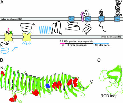
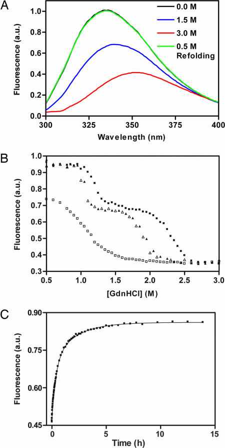
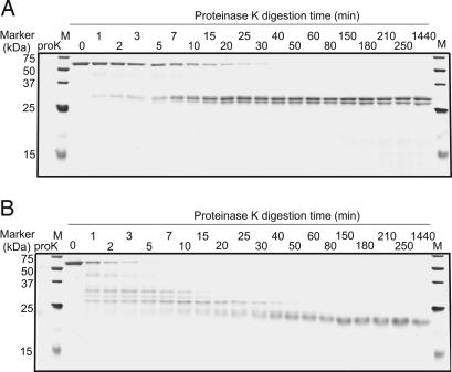
Many virulence factors secreted from pathogenic Gram-negative bacteria are autotransporter proteins. The final step of autotransporter secretion is C --> N-terminal threading of the passenger domain through the outer membrane (OM), mediated by a cotranslated C-terminal porin domain. The native structure is formed only after this final secretion step, which requires neither ATP nor a proton gradient. Sequence analysis reveals that, despite size, sequence, and functional diversity among autotransporter passenger domains, >97% are predicted to form parallel beta-helices, indicating this structural topology may be important for secretion. We report the folding behavior of pertactin, an autotransporter passenger domain from Bordetella pertussis. The pertactin beta-helix folds reversibly in isolation, but folding is much slower than expected based on size and native-state topology. Surprisingly, pertactin is not prone to aggregation during folding, even though folding is extremely slow. Interestingly, equilibrium denaturation results in the formation of a partially folded structure, a stable core comprising the C-terminal half of the protein. Examination of the pertactin crystal structure does not reveal any obvious reason for the enhanced stability of the C terminus. In vivo, slow folding would prevent premature folding of the passenger domain in the periplasm, before OM secretion. Moreover, the extra stability of the C-terminal rungs of the beta-helix might serve as a template for the formation of native protein during OM secretion; hence, vectorial folding of the beta-helix could contribute to the energy-independent translocation mechanism. Coupled with the sequence analysis, the results presented here suggest a general mechanism for autotransporter secretion.


Conflict of interest statement

Conflict of interest statement: No conflicts declared.

Figures

Fig. 1.
Fig. 1.
Pertactin biogenesis and native structure. (A) Current models for AT secretion include passage through the IM via an N-terminal signal sequence (pink), followed by insertion of the 30-kDa C-terminal porin sequence (blue) into the OM; the passenger domain (black) then passes through the central pore of the porin [or a cluster of porins (3)] before folding to the native structure outside the cell. (B) Ribbon diagrams of the pertactin crystal structure (10). Each rung of the parallel β-helix consists of three parallel β-strands arranged in a roughly V-shaped cross section, connected by loops of varying length, producing a long parallel β-sheet on each of the three faces of the β-helix. The locations of the seven Trp residues are represented as red space-filled atoms; Ala-335 is shown in blue. (C) A cross-section view through the seven central rungs of the β-helix (residues 140–357), showing the processively wound stacking of the parallel β-strands and the variable lengths of the connecting loops. The longest connecting loop (35 residues) contains the RGD integrin binding sequence.
Fig. 2.
Fig. 2.
Length distribution of AT proteins, including those predicted to form a right-handed parallel β-helix structure (filled bars) and those predicted not to form a β-helix (open bars).
Fig. 3.
Fig. 3.
Pertactin Trp fluorescence. (A) Emission spectra of native (black), partly folded (blue; in 1.5 M Gdn·HCl), and denatured (red; in 3 M Gdn·HCl) pertactin, and refolded pertactin in 0.5 M Gdn·HCl after unfolding at 4 M Gdn·HCl (green). (B) Pertactin steady-state fluorescence emission at various Gdn·HCl concentrations. Unfolding samples (filled symbols) were incubated for the times shown; refolding samples (open symbols) were first unfolded in 4 M Gdn·HCl for at least 12 h before dilution to the indicated denaturant concentrations. Each time point represents a fresh aliquot from a large sample volume. (C) Time course of pertactin refolding. Pertactin was unfolded for 30 min in 4 M Gdn·HCl and diluted to 0.5 M Gdn·HCl. a.u., arbitrary units.
Fig. 4.
Fig. 4.
Characterization of the pertactin partially folded state. ProK digestion patterns of native pertactin (A) and pertactin equilibrated in 1.5 M Gdn·HCl (B), resolved by SDS/PAGE and silver staining. In both experiments, the pertactin:proK ratio was 2,000:1. Digestion was stopped by boiling for 10 min. As a control for autoproteolysis, proK alone was incubated for 50 min (lane proK). Molecular mass standards are indicated in kDa.
Fig. 5.
Fig. 5.
Mass analysis of the pertactin 21-kDa stable core. (A) MALDI-TOF mass analysis of tryptic peptides from the pertactin 21-kDa stable core fragment. Peaks corresponding to identified tryptic peptides are labeled. No fragments >4,000 m/z were detected. (B) Locations of the tryptic peptides in the full-length pertactin sequence. All labeled peaks deviate less than ±2 Da from the calculated size. Residue numbering is taken from numbering in the PDB file (PDB ID code 1DAB). The upper bar shows the complete preprotein sequence from B. pertussis, including the N-terminal signal sequence, the flexible proline-rich repeat (PRR) at the C terminus of β-helix, and the C-terminal β-porin domain. The middle bar represents the mature β-helix domain used in this study, with labeled tryptic peptides magnified in the lower bar. Symbols correspond to labeled peaks in A.

References

    1. Henderson I. R., Navarro-Garcia F., Desvaux M., Fernandez R. C., Ala’Aldeen D. Microbiol. Mol. Biol. Rev. 2004;68:692–744. - PMC - PubMed
    1. Oomen C. J., Van Ulsen P., Van Gelder P., Feijen M., Tommassen J., Gros P. EMBO J. 2004;23:1257–1266. - PMC - PubMed
    1. Veiga E., Sugawara E., Nikaido H., de Lorenzo V., Fernandez L. A. EMBO J. 2002;21:2122–2131. - PMC - PubMed
    1. Skillman K. M., Barnard T. J., Peterson J. H., Ghirlando R., Bernstein H. D. Mol. Microbiol. 2005;58:945–958. - PubMed
    1. Klauser T., Pohlner J., Meyer T. F. EMBO J. 1990;9:1991–1999. - PMC - PubMed

Publication types

MeSH terms

Associated data