. 2006 Mar 28;103(13):4982-7.
doi: 10.1073/pnas.0505123103. Epub 2006 Mar 20.

TEX14 is essential for intercellular bridges and fertility in male mice

Affiliations

TEX14 is essential for intercellular bridges and fertility in male mice

Michael P Greenbaum et al. Proc Natl Acad Sci U S A. .

Abstract

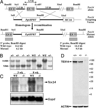
Cytokinesis in somatic cells concludes with the formation of a midbody, which is abscised to form individual daughter cells. In contrast, germ cell cytokinesis results in a permanent intercellular bridge connecting the daughter cells through a large cytoplasmic channel. During spermatogenesis, proposed roles for the intercellular bridge include germ cell communication, synchronization, and chromosome dosage compensation in haploid cells. Although several essential components of the midbody have recently been identified, essential components of the vertebrate germ cell intercellular bridge have until now not been described. Herein, we show that testis-expressed gene 14 (TEX14) is a novel protein that localizes to germ cell intercellular bridges. In the absence of TEX14, intercellular bridges are not observed by using electron microscopy and other markers. Spermatogenesis in Tex14(-/-) mice progresses through the transit amplification of diploid spermatogonia and the expression of early meiotic markers but halts before the completion of the first meiotic division. Thus, TEX14 is required for intercellular bridges in vertebrate germ cells, and these studies provide evidence that the intercellular bridge is essential for spermatogenesis and fertility.


Conflict of interest statement

Conflict of interest statement: No conflicts declared.

Figures

Fig. 1.
Fig. 1.
Targeting of the Tex14 allele and creation of Tex14 mutant mice. (A) A targeting vector was constructed to replace exon 10 of the mouse Tex14 gene with a PgkHPRT expression cassette. The MC1tk expression cassette was used for negative selection. Twenty-two of 70 (31.4%) of the ES cell clones analyzed were correctly targeted at the Tex14 locus. Four targeted ES cell clones were injected into blastocysts to produce 23 chimeric male mice (51, 52), which were bred to produce F1 Tex14+/− offspring. (B) Southern blot analysis of genomic DNA derived from a litter from Tex14+/− (+/−) intercrosses. Similar percentages of male and female mice were genotyped as Tex14 homozygous null (−/−) (see Table 1). (C) Northern blot analysis of RNA derived from WT and Tex14 mutant mice. RNA was probed with 5′ Tex14 or Gapd cDNAs. (D) Western blot analysis of testis samples from 11-day-old Tex14 WT, +/−, and −/− mice by using a polyclonal antibody to TEX14 (Upper) or an antibody to β-actin as a control for sample loading (Lower).
Fig. 2.
Fig. 2.
Gross and histological analysis of postnatal testes. (A) Gross analysis of adult testes from 7-week-old littermate mice. (BI) Histological analysis of testes of 49-day-old Tex14+/− (B and D) and Tex14−/− (C and E) mice, 10-day-old Tex14+/− (F) and Tex14−/− (G) mice, 14-day-old Tex14+/− (H) and Tex14−/− (I) mice, and 21-day-old Tex14+/− (J) and Tex14−/− (K) mice. D, dying spermatocytes; L, leptotene spermatocyte; P, pachytene spermatocyte; PL, preleptotene spermatocyte; Sc, Sertoli cells; Sd6, stage 6 spermatids; Sd15, stage 15 spermatids; Sg-a, type A spermatogonium; Sg-b, type B spermatogonium; V, vacuoles. VI in D designates a stage VI seminiferous tubule. [Scale bars: 200 μm (B), 100 μm (C), 15 μm (D and E), 20 μm (FI), and 40 μm (J and K).]
Fig. 3.
Fig. 3.
Quantitative and qualitative analysis of Tex+/− and Tex14−/− testes. (A) Northern blot analysis of 3-week-old and 8-week-old Tex14+/− and Tex14−/− littermates. (B) Immunohistochemical analysis of 21-day-old Tex14+/− and Tex14−/− testes using antibodies to SYCP3 or cyclin A1 (CCNA1) (arrows point to rare pachytene spermatocytes). (C and D) BrdU labeling (C) and quantitative analysis (D) of BrdU-labeled spermatogonia (Sg) and spermatocytes (Sp) in Tex14+/− (Het) and Tex14−/− (KO) testes. (E) DNA laddering analysis of testes from 10-, 14-, and 56-day-old mice. (F) TUNEL analysis was performed on the testes of 10-day-old and 14-day-old Tex14+/− and Tex14−/− littermates. [Scale bars: 25 μm (B and C) and 50 μm (F).]
Fig. 4.
Fig. 4.
Immunohistological localization of TEX14 to intercellular bridges. (A) Immunofluorescence of a 6-week-old WT seminiferous tubule in which staining for TEX14 (red), actin (green), and DNA (DAPI, blue) were merged. Note the presence of the TEX14-positive (red) rings throughout the tubule. (BD) Immunofluorescence of 9-day-old WT intercellular bridges showing TEX14 (B) and HSF2 (C) and merged with DAPI (D). (E) Immunohistochemistry labeling of TEX14 at intercellular bridges between spermatocytes (black arrows) and spermatogonia (black arrowhead). (F and G) HSF2 immunofluorescence of 9-day-old Tex14-null testis lacking intercellular bridge localization (F) and the corresponding HSF2 protein levels in 9-day-old testes by Western blot analysis (G). [Scale bars: 25 μm (A) and 10 μm (BF).]
Fig. 5.
Fig. 5.
Immunolocalization of TEX14 and PLZF in WT and Tex14−/− mice. TEX14 (red) localizes to the intercellular bridges between PLZF-positive spermatogonia (green) in 8-day-old, neonatal (A) and 8 week-old, mature (B) mice. PLZF labeling of whole-mount seminiferous tubules shows As, Apr, and Aal spermatogonia in 35-day-old Tex14+/− tubules (C), whereas predominantly As spermatogonia appear to be present in 35-day-old Tex14−/− tubules (D). [Scale bar: 10 μm (A and B).]

References

    1. Burgess D. R., Chang F. Trends Cell Biol. 2005;15:156–162. - PubMed
    1. Albertson R., Riggs B., Sullivan W. Trends Cell Biol. 2005;15:92–101. - PubMed
    1. Glotzer M. Science. 2005;307:1735–1739. - PubMed
    1. Skop A. R., Liu H., Yates J., III, Meyer B. J., Heald R. Science. 2004;305:61–66. - PMC - PubMed
    1. Low S. H., Li X., Miura M., Kudo N., Quinones B., Weimbs T. Dev. Cell. 2003;4:753–759. - PubMed

Publication types

Substances