

False positive and false negative FDG-PET scans in various thoracic diseases
- PMID: 16549957
- PMCID: PMC2667579
- DOI: 10.3348/kjr.2006.7.1.57
False positive and false negative FDG-PET scans in various thoracic diseases
Abstract
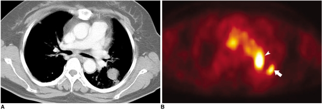
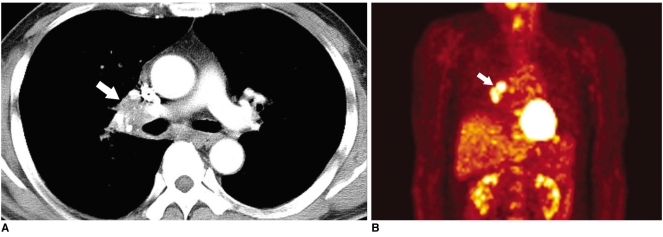
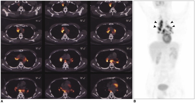
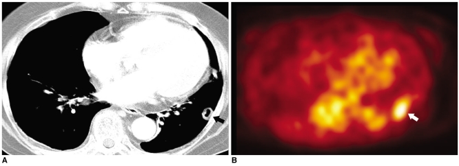
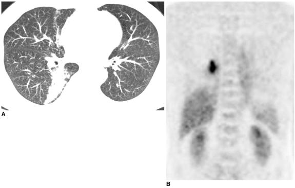
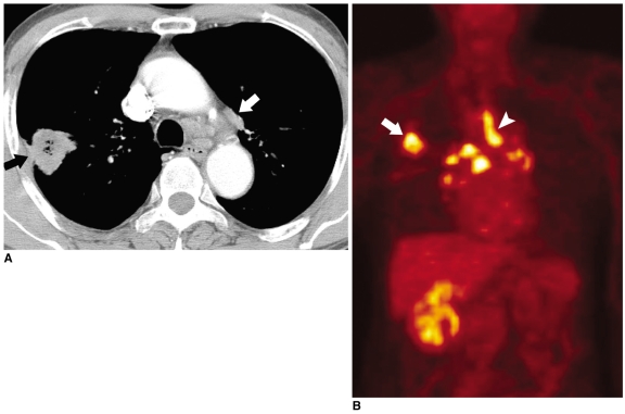
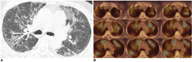
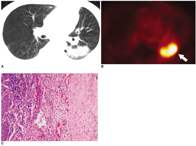
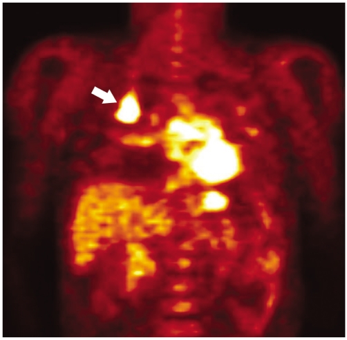
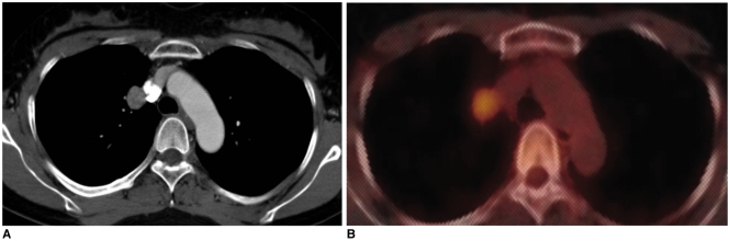
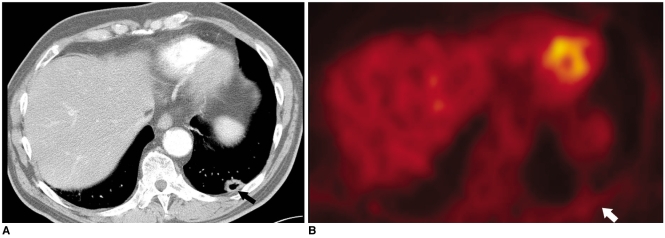
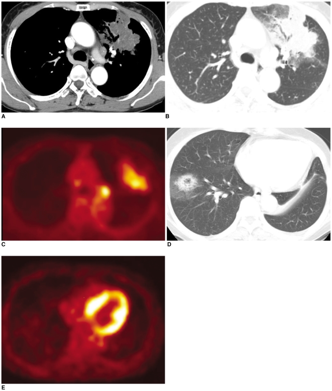
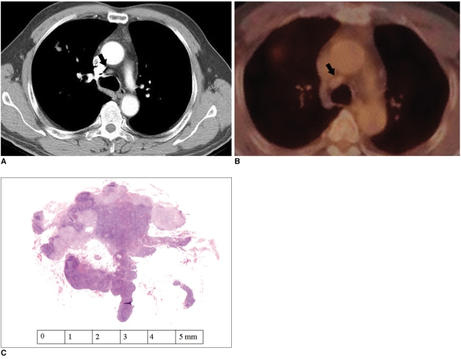
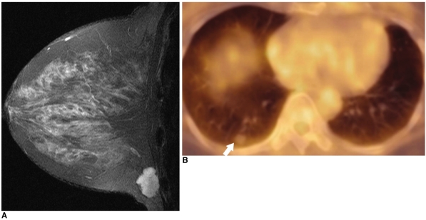
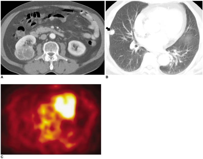
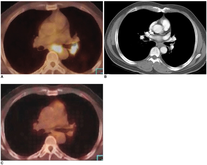
Fluorodeoxyglucose (FDG)-positron emission tomography (PET) is being used more and more to differentiate benign from malignant focal lesions and it has been shown to be more efficacious than conventional chest computed tomography (CT). However, FDG is not a cancer-specific agent, and false positive findings in benign diseases have been reported. Infectious diseases (mycobacterial, fungal, bacterial infection), sarcoidosis, radiation pneumonitis and post-operative surgical conditions have shown intense uptake on PET scan. On the other hand, tumors with low glycolytic activity such as adenomas, bronchioloalveolar carcinomas, carcinoid tumors, low grade lymphomas and small sized tumors have revealed false negative findings on PET scan. Furthermore, in diseases located near the physiologic uptake sites (heart, bladder, kidney, and liver), FDG-PET should be complemented with other imaging modalities to confirm results and to minimize false negative findings. Familiarity with these false positive and negative findings will help radiologists interpret PET scans more accurately and also will help to determine the significance of the findings. In this review, we illustrate false positive and negative findings of PET scan in a variety of diseases.
Figures
References
-
- Goo JM, Im JG, Do KH, Yeo JS, Seo JB, Kim HY, et al. Pulmonary tuberculoma evaluated by means of FDG PET: findings in 10 cases. Radiology. 2000;216:117–121. - PubMed
-
- Kostakoglu L, Agress H, Goldsmith SJ. Clinical role of FDG PET in evalutaion of cancer patients. RadioGraphics. 2003;23:315–340. - PubMed
-
- Trukington TG, Coleman RE. Clinical oncologic PET: An Introduction. Semin Roentgenol. 2002;37:102–109. - PubMed
-
- Gupta NC, Graeber GM, Bishop HA. Comparative efficacy of positron emission tomography with fluorodeoxyglucose in evaluation of small (< 1 cm), intermediate (1 to 3 cm), and large (> 3 cm) lymph node lesions. Chest. 2000;117:773–778. - PubMed
-
- Alavi A, Gupta N, Alberini JL, Hickeson M, Adam LE, Bhargava P, et al. Positron emission tomography Imaging in nonmalignant thoracic disorders. Semin Nucl Med. 2002;32:293–321. - PubMed
Publication types
MeSH terms
Substances
LinkOut - more resources
Full Text Sources
Other Literature Sources
Medical
