. 2006 Mar 21;7(1):39.
doi: 10.1186/1465-9921-7-39.

IL-4 induced MUC4 enhancement in respiratory epithelial cells in vitro is mediated through JAK-3 selective signaling

Affiliations

IL-4 induced MUC4 enhancement in respiratory epithelial cells in vitro is mediated through JAK-3 selective signaling

Gautam Damera et al. Respir Res. .

Abstract

Background: Recent studies have identified MUC4 mucin as a ligand for activation of ErbB2, a receptor tyrosine kinase that modulates epithelial cell proliferation following epithelial damage in airways of asthmatics. In this study, we investigated the potential role of IL-4, one of the Th2 inflammatory cytokines persistent in asthmatic airways, in regulating MUC4 expression using a cell line NCI-H650.

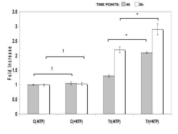
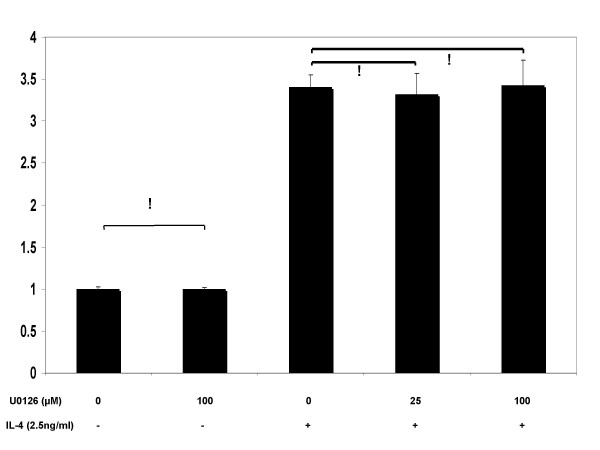
Methods: Real time PCR analysis was performed to determine concentration and time dependent effects of IL-4 upon MUC4 expression. Nuclear run on experiments were carried out to explore potential transcriptional modulation. Western blotting experiments using a monoclonal antibody specific to ASGP-2 domain of MUC4 were performed to analyze MUC4 glycoprotein levels in plasma membrane fractions. To analyze potential signal transduction cascades, IL-4 treated confluent cultures were co-incubated, separately with a pan-JAK inhibitor, a JAK-3 selective inhibitor or a MEK-1, 2 (MAPK) inhibitor at various concentrations before MUC4 transcript analysis. Corresponding transcription factor activation was tested by western blotting using a monoclonal p-STAT-6 antibody.

Results: MUC4 levels increased in a concentration and time specific fashion reaching peak expression at 2.5 ng/ml and 8 h. Nuclear run on experiments revealed transcriptional enhancement. Corresponding increases in MUC4 glycoprotein levels were observed in plasma membrane fractions. Pan-JAK inhibitor revealed marked reduction in IL-4 stimulated MUC4 levels and JAK3 selective inhibitor down-regulated MUC4 mRNA expression in a concentration-dependent fashion. In accordance with the above observations, STAT-6 activation was detected within 5 minutes of IL-4 stimulus. No effect in MUC4 levels was observed on using a MAPK inhibitor.

Conclusion: These observations signify a potential role for IL-4 in MUC4 up-regulation in airway epithelia.


Figures

Figure 1
Figure 1
(a) RT-PCR analysis of IL-4R mRNA expression from NCI-H650 cells. Total RNA was extracted from confluent cultures and analyzed for the presence of IL-4R and GAPDH transcripts by RT-PCR. The amplified products were run on 1% agarose-ethidium bromide gels. Bands at 335 bp (IL-4R) and 1000 bp (GAPDH) were observed. Immunohistochemical determination of IL-4Rα upon permeabilized NCI-H650 cells using (b) rabbit polyclonal anti-human IL-4Rα antibody (c) and non-immune rabbit IgG serum (control). Secondary antibody incubations were performed with Alexa Fluor (568 nm)-labeled mouse antibody. Cells were counterstained with DAPI and photographed using dapi: rhodamine filters at 25:2000 ms exposure. Block arrows in (1b) are indicative of specific staining to IL-4 receptors on cell surface.
Figure 2
Figure 2
Dose-dependent expression of MUC4 by IL-4. NCI-H650 cells were treated with 0 to 10 ng/ml concentrations of IL-4. The mRNA levels of MUC4 were determined by real-time PCR analysis. Data indicated in the graph are mean fold increase ± SE over mean control value. The data are representative of three independent experiments with each treatment run in triplicate. * significantly different, p < 0.05.
Figure 3
Figure 3
Time course of MUC4 gene expression by IL-4. NCI-H650 cells were treated with 2.5 ng/ml of IL-4 for indicated times. Controls were sham treated. [▫] MUC4 and [▪] 18S rRNA eukaryotic mRNA levels were determined by real-time PCR amplification. Data indicated in the graph are mean fold increase ± SE over mean control value. The graph summarizes data from three independent experiments with each treatment run in triplicate. * significantly different, p < 0.05.
Figure 4
Figure 4
Transcriptional regulation of MUC4 by IL-4. Nuclei were isolated from IL-4 treated and control cells at two separate time points of 4 h and 8 h. The extracted nuclei were incubated with or without a mixture of NTP's (0.5 mM each) for 45 min. Real-time PCR amplifications were performed on total RNA extracted from: C(-NTP), untreated nuclei of control cells; C(+NTP), NTP treated nuclei of control cells; Tr(-NTP), untreated nuclei from IL-4-treated cells; and Tr(+NTP), NTP treated nuclei from IL-4-treated cells. Data indicated in the graph are mean fold increase ± SE over mean control value. The graph summarizes data from three independent experiments with triplicate samples. * significant increase (p < 0.05) ; ! no significant difference (p > 005)
Figure 5
Figure 5
MUC4 glycoprotein expression on IL-4 stimulus. Panel (A): Confluent cells were treated with (Tr) 2.5 ng/ml of IL-4 for 8 h followed by a 16 h chase period in fresh medium without IL-4. (Ct) Untreated control cells. Plasma membrane protein (50 μg) was resolved on 4–20% SDS-PAGE gradient gels and evaluated with 1G8 anti-MUC4 monoclonal primary antibody and horse radish peroxidase conjugated goat secondary antibody. The Western blots are representative of three separate experiments. Panel (B): Commassie blue staining of the gels.
Figure 6
Figure 6
Effects of pan-JAK signaling inhibitor (DBI), upon IL-4-stimulated MUC4 mRNA expression. NCI-H650 cells were pretreated with indicated concentrations of DBI for 30 min before stimulation with 2.5 ng/ml of IL-4. Data indicated in the graph are mean fold increase ± SE over mean control value. The graph summarizes data from three independent experiments with triplicate samples. * significantly different (p < 0.05), ! no statistical significance, (p > 0.05).
Figure 7
Figure 7
Effects of JAK-3 selective signaling inhibitor (WHI-P131), upon IL-4-stimulated MUC4 mRNA expression. NCI-H650 cells were pretreated with indicated concentrations of WHI-P131 for 30 min before stimulation with 2.5 ng/ml of IL-4. Data indicated in the graph are mean fold increase ± SE over mean control value. The graph summarizes data from three independent experiments with triplicate samples. * significantly different (p < 0.05), ! no statistical significance, (p > 0.05).
Figure 8
Figure 8
Effects of MAPK signaling inhibitor (U0126), upon IL-4-stimulated MUC4 mRNA expression. NCI-H650 cells were pretreated with indicated concentrations of U0126 for 30 min before stimulation with 2.5 ng/ml of IL-4. Data indicated in the graph are mean fold increase ± SE over mean control value. The graph summarizes data from three independent experiments with triplicate samples. ! no statistical significance, (p > 0.05).
Figure 9
Figure 9
Activation of STAT-6 by IL-4 in NCI-H650 cells. Western analysis of cell lysates from 2.5 ng/ml IL-4 stimulated cells at indicated time points. STAT-6 activation was detected using an anti-phospho-STAT-6 antibody. The Western blots are representative of three separate experiments with triplicate samples.

References

    1. Kay AB. Asthma and inflammation. J Allergy Clin Immunol. 1991;87:893–910. doi: 10.1016/0091-6749(91)90408-G. - DOI - PubMed
    1. Fahy JV. Remodeling of the airway epithelium in asthma. Am J Respir Crit Care Med. 2001;164:S46–51. - PubMed
    1. Laitinen LA, Heino M, Laitinen A, Kava T, Haahtela T. Damage of the airway epithelium and bronchial reactivity in patients with asthma. Am Rev Respir Dis. 1985;131:599–606. - PubMed
    1. Frew AJ, Moqbel R, Azzawi M, Hartnell A, Barkans J, Jeffery PK, Kay AB, Scheper RJ, Varley J, Church MK, et al. T lymphocytes and eosinophils in allergen-induced late-phase asthmatic reactions in the guinea pig. Am Rev Respir Dis. 1990;141:407–413. - PubMed
    1. Azzawi M, Bradley B, Jeffery PK, Frew AJ, Wardlaw AJ, Knowles G, Assoufi B, Collins JV, Durham S, Kay AB. Identification of activated T lymphocytes and eosinophils in bronchial biopsies in stable atopic asthma. Am Rev Respir Dis. 1990;142:1407–1413. - PubMed

Publication types

MeSH terms