Central nervous system extraosseous Ewing sarcoma: radiologic manifestations of this newly defined pathologic entity
- PMID: 16551995
- PMCID: PMC7976978
Central nervous system extraosseous Ewing sarcoma: radiologic manifestations of this newly defined pathologic entity
Abstract
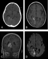
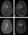
Although these entities are histologically similar, recent advances in molecular genetics have allowed the distinction of central nervous system extraosseous Ewing sarcoma (CNS-EES) from central primitive neuroectodermal tumors (c-PNET) including medulloblastoma and supratentorial PNET. We present 2 cases of pathologically confirmed CNS-EES. Knowledge of CNS-EES as a distinct entity enables the neuroradiologist to suggest the proper diagnosis and the need for special immuno-histochemical and molecular studies to confirm the diagnosis. Because treatment and prognosis are vastly different, the proper diagnosis of CNS-EES versus c-PNET is critical.
Figures


References
-
- Tefft M, Vawter GF, Mitus A. Paravertebral “round cell” tumors in children. Radiology 1969;92:1501–09 - PubMed
-
- Ahmad R, Mayol B, Davis M, et al. Extraskeletal Ewing’s sarcoma. Cancer 1999;85:725–31 - PubMed
-
- Kovar H. Ewing’s sarcoma and peripheral primitive neuroectodermal tumors after their genetic union. Curr Opin Oncol 1998;10:334–42 - PubMed
-
- Schmidt D, Hermann C, Jurgens H, et al. Malignant peripheral neuroectodermal tumor and its necessary distinction from Ewing’s sarcoma: a report from Kiel pediatric tumor registry. Cancer 1991;68:2251–59 - PubMed
-
- Dirks PB, Harris L, Hoffman HJ, et al. Supratentorial primitive neuroectodermal tumors in children. J Neurooncol 1996;29:75–84 - PubMed
Publication types
MeSH terms
LinkOut - more resources
Full Text Sources
Medical
