

Imaging and nanomedicine for diagnosis and therapy in the central nervous system: report of the eleventh annual Blood-Brain Barrier Disruption Consortium meeting
- PMID: 16552023
- PMCID: PMC7976977
Imaging and nanomedicine for diagnosis and therapy in the central nervous system: report of the eleventh annual Blood-Brain Barrier Disruption Consortium meeting
Abstract
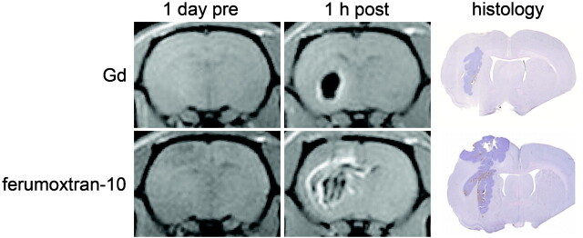
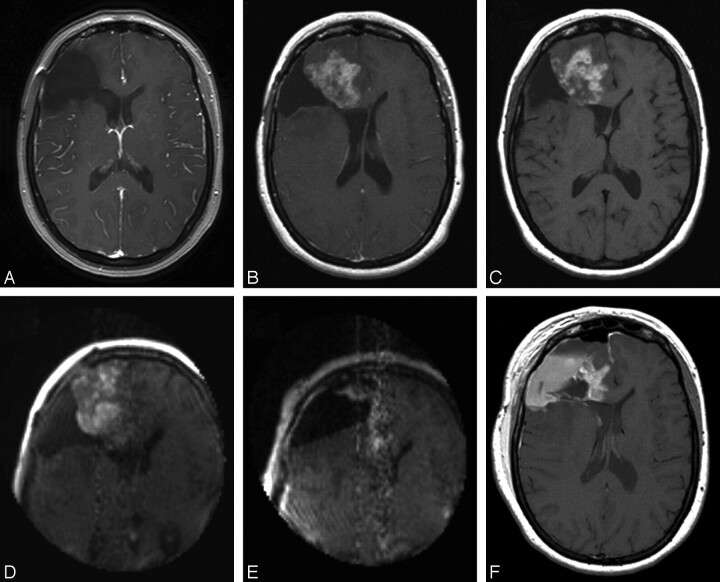
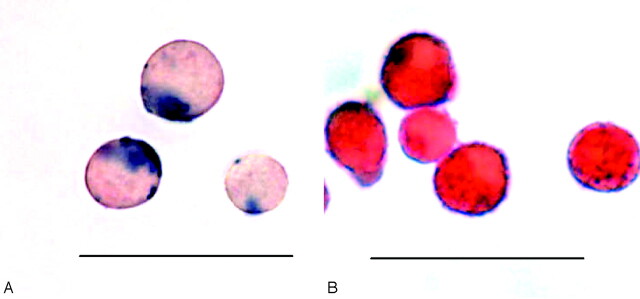
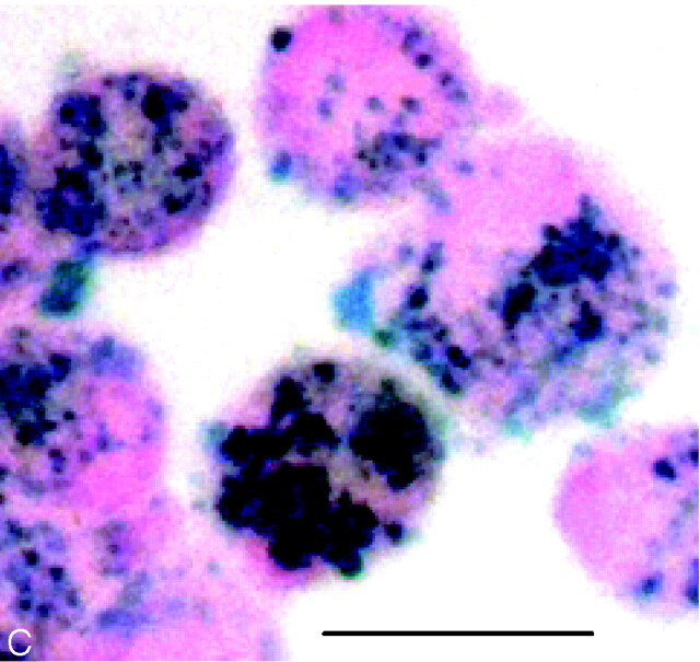
The blood-brain barrier (BBB) presents a major obstacle to the treatment of malignant brain tumors and other central nervous system (CNS) diseases. The Eleventh Annual Blood-Brain Barrier Disruption Consortium Meeting was convened to discuss recent advances and future directions in imaging and nanomedicine. Two sessions, one on Cell and Molecular Imaging in the CNS and another on Nanotechnology, Nanobiology, and Nanomedicine, were held March 17-18, 2005, in Portland, Ore. CNS imaging presentations targeted differentiating tumor, neural lesions, and necrosis from healthy brain tissue; methods of delivery of imaging agents across the BBB; and new iron oxide-based nanoparticle contrast agents for MR imaging. Nanobiology presentations covered the development of new nanotechnology and its use in imaging, diagnosis, and therapy in the CNS. Discussions at this meeting stressed the role of biotechnology in the convergence of CNS imaging and nanomedicine and are summarized in this article.
Figures
Similar articles
-
New frontiers in translational research in neuro-oncology and the blood-brain barrier: report of the tenth annual Blood-Brain Barrier Disruption Consortium Meeting.Clin Cancer Res. 2005 Jan 15;11(2 Pt 1):421-8. Clin Cancer Res. 2005. PMID: 15701824
-
Advances in nanomedicines for diagnosis of central nervous system disorders.Biomaterials. 2021 Feb;269:120492. doi: 10.1016/j.biomaterials.2020.120492. Epub 2020 Oct 24. Biomaterials. 2021. PMID: 33153757 Review.
-
Delivery of chemotherapy and antibodies across the blood-brain barrier and the role of chemoprotection, in primary and metastatic brain tumors: report of the Eleventh Annual Blood-Brain Barrier Consortium meeting.J Neurooncol. 2007 Jan;81(1):81-91. doi: 10.1007/s11060-006-9209-y. Epub 2006 Jul 21. J Neurooncol. 2007. PMID: 16858513
-
Application of Therapeutic Nanoplatforms as a Potential Candidate for the Treatment of CNS Disorders: Challenges and Possibilities.Curr Pharm Des. 2022;28(33):2742-2757. doi: 10.2174/1381612828666220729104433. Curr Pharm Des. 2022. PMID: 35909283 Review.
-
Role of Nanomedicine-Based Therapeutics in the Treatment of CNS Disorders.Molecules. 2023 Jan 28;28(3):1283. doi: 10.3390/molecules28031283. Molecules. 2023. PMID: 36770950 Free PMC article. Review.
Cited by
-
"Extremely minimally invasive": recent advances in nanotechnology research and future applications in neurosurgery.Neurosurg Rev. 2015 Jan;38(1):27-37; discussion 37. doi: 10.1007/s10143-014-0566-2. Epub 2014 Aug 31. Neurosurg Rev. 2015. PMID: 25173621 Review.
-
Superparamagnetic iron oxide nanoparticles: diagnostic magnetic resonance imaging and potential therapeutic applications in neurooncology and central nervous system inflammatory pathologies, a review.J Cereb Blood Flow Metab. 2010 Jan;30(1):15-35. doi: 10.1038/jcbfm.2009.192. Epub 2009 Sep 16. J Cereb Blood Flow Metab. 2010. PMID: 19756021 Free PMC article. Review.
-
Effect of Conducting, Semi-Conducting and Insulating Nanoparticles on AC Breakdown Voltage and Partial Discharge Activity of Synthetic Ester: A Statistical Analysis.Nanomaterials (Basel). 2022 Jun 19;12(12):2105. doi: 10.3390/nano12122105. Nanomaterials (Basel). 2022. PMID: 35745444 Free PMC article.
-
Comparative analysis of ferumoxytol and gadoteridol enhancement using T1- and T2-weighted MRI in neuroimaging.AJR Am J Roentgenol. 2011 Oct;197(4):981-8. doi: 10.2214/AJR.10.5992. AJR Am J Roentgenol. 2011. PMID: 21940589 Free PMC article.
-
Tracking Neural Progenitor Cell Migration in the Rodent Brain Using Magnetic Resonance Imaging.Front Neurosci. 2019 Jan 11;12:995. doi: 10.3389/fnins.2018.00995. eCollection 2018. Front Neurosci. 2019. PMID: 30686969 Free PMC article. Review.
References
-
- Delbeke D, Meyerowitz C, Lapidus RL, et al. Optimal cutoff levels of F-18 fluorodeoxyglucose uptake in the differentiation of low-grade from high-grade brain tumors with PET. Radiology 1995;195:47–52 - PubMed
-
- Spence AM, Muzi M, Graham MM, et al. 2-[(18)F]Fluoro-2-deoxyglucose and glucose uptake in malignant gliomas before and after radiotherapy: correlation with outcome. Clin Cancer Res 2002;8:971–79 - PubMed
-
- Degen JW, Walbridge S, Vortmeyer AO, et al. Safety and efficacy of convection-enhanced delivery of gemcitabine or carboplatin in a malignant glioma model in rats. J Neurosurg 2003;99:893–98 - PubMed
-
- Parney IF, Kunwar S, McDermott M, et al. Neuroradiographic changes following convection-enhanced delivery of the recombinant cytotoxin interleukin 13-PE38QQR for recurrent malignant glioma. J Neurosurg 2005;102:267–75 - PubMed
Publication types
MeSH terms
Grants and funding
LinkOut - more resources
Full Text Sources