. 2006 Apr;74(4):2052-62.
doi: 10.1128/IAI.74.4.2052-2062.2006.

Analysis of involvement of the RecF pathway in p44 recombination in Anaplasma phagocytophilum and in Escherichia coli by using a plasmid carrying the p44 expression and p44 donor loci

Affiliations

Analysis of involvement of the RecF pathway in p44 recombination in Anaplasma phagocytophilum and in Escherichia coli by using a plasmid carrying the p44 expression and p44 donor loci

Quan Lin et al. Infect Immun. 2006 Apr.

Abstract

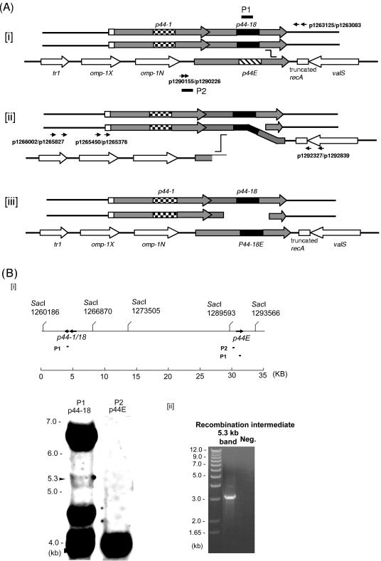
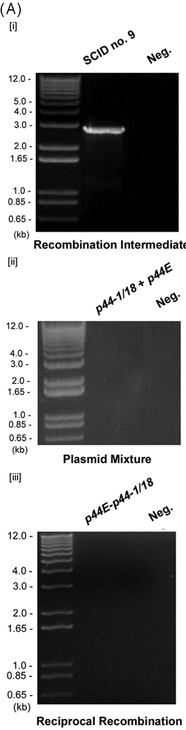
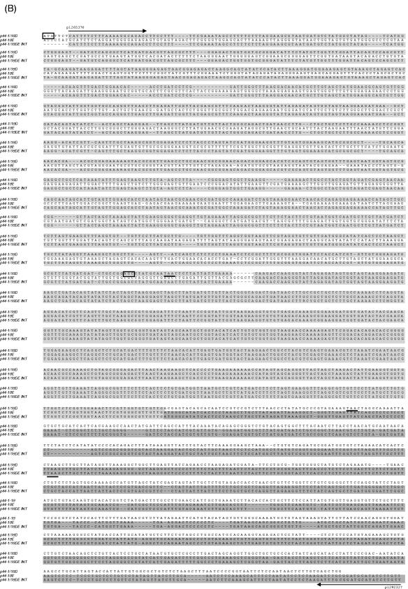
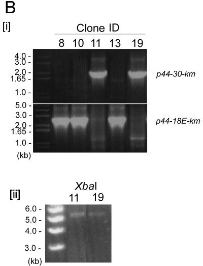
Anaplasma phagocytophilum, the etiologic agent of human granulocytic anaplasmosis, has a large paralog cluster (approximate 90 members) that encodes the 44-kDa major outer membrane proteins (P44s). Gene conversion at a single p44 expression locus leads to P44 antigenic variation. Homologs of genes for the RecA-dependent RecF pathway, but not the RecBCD or RecE pathways, of recombination were detected in the A. phagocytophilum genome. In the present study, we examined whether the RecF pathway is involved in p44 gene conversion. The recombination intermediate structure between a donor p44 and the p44 expression locus of A. phagocytophilum was detected in an HL-60 cell culture by Southern blot analysis followed by sequencing the band and in blood samples from infected SCID mice by PCR, followed by sequencing. The sequences were consistent with the RecF pathway recombination: a half-crossover structure, consisting of the donor p44 locus connected to the 3' conserved region of the recipient p44 in the p44 expression locus in direct orientation. To determine whether the p44 recombination intermediate structure can be generated in a RecF-active Escherichia coli strain, we constructed a double-origin plasmid carrying the p44 expression locus and a donor p44 locus and introduced the plasmid into various E. coli strains. The recombination intermediate was recovered in an E. coli strain with active RecF recombination pathway but not in strains with deficient RecF pathway. Our results support the view that the p44 gene conversion in A. phagocytophilum occurs through the RecF pathway.


Figures

FIG. 1.
FIG. 1.
Analysis of the p44 recombination intermediate in A. phagocytophilum. (A) Successive half-crossover model for p44-18 conversion to the p44E and the experimental design. [i] The donor locus was expected to be a part of replicated chromosome; the p44-1/18 locus in a donor chromosome (p44-1/18D) synapses with the 3′ conserved region of p44E in the recipient chromosome. [ii] A half crossover between p44E and p44-1/18D generates one recombination duplex and two ends. The upstream of the synapsed region of p44E is presumably degraded to generate a single-stranded tail at the 5′ conserved region by an exonuclease. [iii] The putative final products of the successive crossover are one duplex with two double strands exchanged in the recipient p44 expression locus and two ends in the donor locus. The gray boxes are p44 conserved regions, and the large checkerboard, black, and wide downward diagonal are hypervariable regions of p44-1, p44-18, and the recipient p44E, respectively. The arrows of boxes indicate transcriptional orientation. Horizontal arrows with numbers indicate primers used to isolate the intermediate structure by PCR and primers used to demonstrate the absence of a reciprocal recombination product. Black short bars indicate the locations of two probes (P1 and P2) used for Southern blot analysis in Fig. 1B. The “stairstep” symbol indicates the potential pair of the recombination sites. (B) Southern blot analysis of the half-crossover intermediate structure of A. phagocytophilum cultured in HL-60 cells. [i] The probe positions and SacI digestion sites within the 35-kb region containing p44-1/18 and p44E are indicated. The numbers under the SacI sites indicate the positions of the cleavage sites in the genome. The genomic DNA was purified from A. phagocytophilum cultivated in HL-60 cells. Southern blot analysis of genomic DNA digested with the restriction enzyme SacI with two probes (P1 and P2) showed the ∼6.6-kb band containing the donor p44-1/18 locus and a 4-kb band containing the recipient p44E. An ∼5.3-kb band contained duplicated p44-1/18. [ii] PCR was performed using the primer pairs p1265450-p1292839 and p1265376-p1292327 (Fig. 1Aii) and the DNA isolated from the 5.3-kb region of the gel as a template. The numbers on the left indicate the molecular sizes. A band of approximately 3.2 kb was amplified and sequenced GenBank accession no. DQ011270. “Neg.” refers to a negative control with water as a template.
FIG.2.
FIG.2.
Analysis of the p44 recombination intermediate in A. phagocytophilum from an infected SCID mouse. (A) The half-crossover recombination intermediate was detected by PCR of peripheral blood leukocytes from an A. phagocytophilum-infected SCID mouse. [i] The ∼2.9-kb band was amplified by the primer pairs p1265450-p1292839 and p1265376-p1292327 (Fig. 1Aii) using Pfu DNA polymerase. [ii] No PCR product was detected with the same primer pairs from uninfected SCID mouse DNA spiked with plasmids containing p44-1/18 and p44E as a template. [iii] No PCR product was detected in peripheral blood leukocytes of infected mice with forward primers located in the intergenic region of p44E and omp-1N and with reverse primers located downstream of donor p44-1/18 locus (Fig. 1Ai). “Neg.” refers to a negative control with water as a template. (B) The nucleotide sequence of the half-crossover recombination duplex between p44E and p44-1/18D. The second set of primers used for the nested PCR are indicated by horizontal arrows and labeled with primer ID. p44-1/18DE INT is the intermediate structure containing donor site p44-1/18 and the recipient site p44E. The sequence is identical to that expected for a half-crossover recombination intermediate as illustrated in Fig. 1Aii. The light-shaded areas indicate sequence identity between the crossover structure and the putative donor p44-1/18 locus, and the dark shaded areas indicate identical sequence between the hybrid structure and the recipient p44E, or among all three structures. The boxed nucleotides are start codons for p44-1 and for p44-18. The stop codons are underlined. Dashes indicate sequence gaps that helped to determine the origins of conserved sequences in the recombination intermediate.
FIG.2.
FIG.2.
Analysis of the p44 recombination intermediate in A. phagocytophilum from an infected SCID mouse. (A) The half-crossover recombination intermediate was detected by PCR of peripheral blood leukocytes from an A. phagocytophilum-infected SCID mouse. [i] The ∼2.9-kb band was amplified by the primer pairs p1265450-p1292839 and p1265376-p1292327 (Fig. 1Aii) using Pfu DNA polymerase. [ii] No PCR product was detected with the same primer pairs from uninfected SCID mouse DNA spiked with plasmids containing p44-1/18 and p44E as a template. [iii] No PCR product was detected in peripheral blood leukocytes of infected mice with forward primers located in the intergenic region of p44E and omp-1N and with reverse primers located downstream of donor p44-1/18 locus (Fig. 1Ai). “Neg.” refers to a negative control with water as a template. (B) The nucleotide sequence of the half-crossover recombination duplex between p44E and p44-1/18D. The second set of primers used for the nested PCR are indicated by horizontal arrows and labeled with primer ID. p44-1/18DE INT is the intermediate structure containing donor site p44-1/18 and the recipient site p44E. The sequence is identical to that expected for a half-crossover recombination intermediate as illustrated in Fig. 1Aii. The light-shaded areas indicate sequence identity between the crossover structure and the putative donor p44-1/18 locus, and the dark shaded areas indicate identical sequence between the hybrid structure and the recipient p44E, or among all three structures. The boxed nucleotides are start codons for p44-1 and for p44-18. The stop codons are underlined. Dashes indicate sequence gaps that helped to determine the origins of conserved sequences in the recombination intermediate.
FIG.3.
FIG.3.
Analysis of a p44 recombination intermediate in E. coli using plasmid encoding p44 expression and donor loci. (A) Double-origin plasmid pEKD30 and E. coli system design. [i] Plasmid pEKD30 carries the recipient site of the p44 expression locus p44E upstream region (p44E IR)- p44-18E-km-p44E downstream sequence (p44E DS) and the donor site p44-30PrecA in a direct orientation. The donor p44-30PrecA has an E. coli recA gene promoter in the hypervariable region (bent arrow). The plasmid carries two more antibiotic-resistant genes (cm and amp) and two compatible replication origins (P15A and ColE). The restriction enzyme cleavage sites are shown. If p44-30PrecA recombines to p44-18E, km is transcribed from the recA promoter, allowing isolation of the recombination intermediates in the presence of Km. [ii] Experimental design. If nonreciprocal recombination occurs between p44-30PrecA and p44-18E, only one type of plasmid (5.7 kb) that carries km with upstream p44-30PrecA is expected to be isolated in the presence of Km. If reciprocal recombination occurs between the donor and the recipient sequences, two plasmids should be generated: one identical to the plasmid generated by the nonreciprocal recombination (5.7 kb) and another that carries p44-18E and amp (3.6 kb). Thus, in nonreciprocal recombination, Kmr E. coli strains are Ams, while in reciprocal recombination Kmr E. coli strains are Amr. [iii] Sequence alignment of 5′- and 3′-end conserved regions of donor p44-30D and recipient p44-18E in the plasmid pEKD30. The different nucleotides between p44-30D and p44-18E conserved regions are shaded in light gray. (B) Analysis of the Kmr Ams plasmids isolated from E. coli strain JC7623 with an active RecF pathway. [i] PCR amplification using the primer pair located upstream of p44-30PrecA and downstream of km, respectively, showed a band of the expected size for the recombined p44-30PrecA-km structure in Kmr Ams clones 11 and 19. The remaining three Kmr Ams clones had the original p44-18-km structure. [ii] The two PCR positive plasmids from clones 11 and 19 were analyzed by digestion with XbaI to determine the size of the recombined plasmid. As predicted from the restriction sites shown in Fig. 3Ai, XbaI digestion generated a single 5.7-kb band from the recombined p44-30-km plasmid. [iii] The sequence between the two SalI sites in clones 11 and 19, which confirmed the recombination between p44-30PrecA and p44-18E.
FIG.3.
FIG.3.
Analysis of a p44 recombination intermediate in E. coli using plasmid encoding p44 expression and donor loci. (A) Double-origin plasmid pEKD30 and E. coli system design. [i] Plasmid pEKD30 carries the recipient site of the p44 expression locus p44E upstream region (p44E IR)- p44-18E-km-p44E downstream sequence (p44E DS) and the donor site p44-30PrecA in a direct orientation. The donor p44-30PrecA has an E. coli recA gene promoter in the hypervariable region (bent arrow). The plasmid carries two more antibiotic-resistant genes (cm and amp) and two compatible replication origins (P15A and ColE). The restriction enzyme cleavage sites are shown. If p44-30PrecA recombines to p44-18E, km is transcribed from the recA promoter, allowing isolation of the recombination intermediates in the presence of Km. [ii] Experimental design. If nonreciprocal recombination occurs between p44-30PrecA and p44-18E, only one type of plasmid (5.7 kb) that carries km with upstream p44-30PrecA is expected to be isolated in the presence of Km. If reciprocal recombination occurs between the donor and the recipient sequences, two plasmids should be generated: one identical to the plasmid generated by the nonreciprocal recombination (5.7 kb) and another that carries p44-18E and amp (3.6 kb). Thus, in nonreciprocal recombination, Kmr E. coli strains are Ams, while in reciprocal recombination Kmr E. coli strains are Amr. [iii] Sequence alignment of 5′- and 3′-end conserved regions of donor p44-30D and recipient p44-18E in the plasmid pEKD30. The different nucleotides between p44-30D and p44-18E conserved regions are shaded in light gray. (B) Analysis of the Kmr Ams plasmids isolated from E. coli strain JC7623 with an active RecF pathway. [i] PCR amplification using the primer pair located upstream of p44-30PrecA and downstream of km, respectively, showed a band of the expected size for the recombined p44-30PrecA-km structure in Kmr Ams clones 11 and 19. The remaining three Kmr Ams clones had the original p44-18-km structure. [ii] The two PCR positive plasmids from clones 11 and 19 were analyzed by digestion with XbaI to determine the size of the recombined plasmid. As predicted from the restriction sites shown in Fig. 3Ai, XbaI digestion generated a single 5.7-kb band from the recombined p44-30-km plasmid. [iii] The sequence between the two SalI sites in clones 11 and 19, which confirmed the recombination between p44-30PrecA and p44-18E.

Similar articles

Cited by

References

    1. Allers, T., and M. Lichten. 2001. Differential timing and control of noncrossover and crossover recombination during meiosis. Cell 106:47-57. - PubMed
    1. Bakken, J. S., J. Krueth, C. Wilson-Nordskog, R. L. Tilden, K. Asanovich, and J. S. Dumler. 1996. Clinical and laboratory characteristics of human granulocytic ehrlichiosis. JAMA 275:199-205. - PubMed
    1. Barbet, A. F., A. Lundgren, J. Yi, F. R. Rurangirwa, and G. H. Palmer. 2000. Antigenic variation of Anaplasma marginale by expression of MSP2 mosaics. Infect. Immun. 68:6133-6138. - PMC - PubMed
    1. Barbet, A. F., P. F. M. Meeus, M. Belanger, M. V. Bowie, J. Yi, A. M. Lundgren, A. R. Alleman, S. J. Wong, F. K. Chu, U. G. Munderloh, and S. D. Jauron. 2003. Expression of multiple outer membrane protein sequence variants from a single genomic locus of Anaplasma phagocytophilum. Infect. Immun. 71:1706-1718. - PMC - PubMed
    1. Barbour, A. G., and B. I. Restrepo. 2000. Antigenic variation in vector-borne pathogens. Emerg. Infect. Dis. 6:449-457. - PMC - PubMed

Publication types

MeSH terms

Substances

Associated data

LinkOut - more resources