Review
. 2006 Aug;291(2):F271-81.
doi: 10.1152/ajprenal.00071.2006. Epub 2006 Mar 22.

Hypoxia-inducible factors in the kidney

Affiliations
Review

Hypoxia-inducible factors in the kidney

Volker H Haase. Am J Physiol Renal Physiol. 2006 Aug.

Abstract

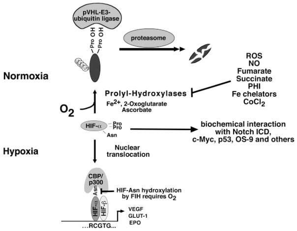
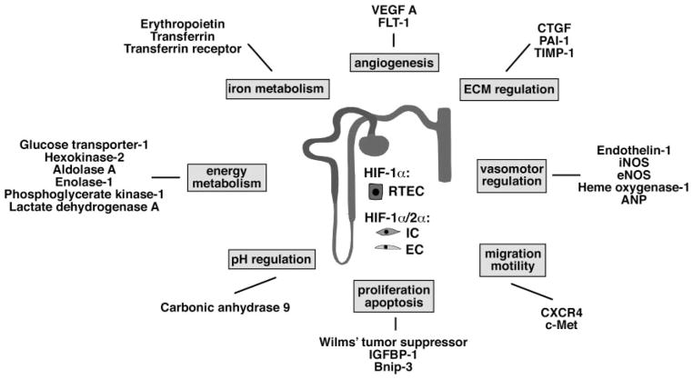
Tissue hypoxia not only occurs under pathological conditions but is also an important microenvironmental factor that is critical for normal embryonic development. Hypoxia-inducible factors HIF-1 and HIF-2 are oxygen-sensitive basic helix-loop-helix transcription factors, which regulate biological processes that facilitate both oxygen delivery and cellular adaptation to oxygen deprivation. HIFs consist of an oxygen-sensitive alpha-subunit, HIF-alpha, and a constitutively expressed beta-subunit, HIF-beta, and regulate the expression of genes that are involved in energy metabolism, angiogenesis, erythropoiesis and iron metabolism, cell proliferation, apoptosis, and other biological processes. Under conditions of normal Po(2), HIF-alpha is hydroxylated and targeted for rapid proteasomal degradation by the von Hippel-Lindau (VHL) E3-ubiquitin ligase. When cells experience hypoxia, HIF-alpha is stabilized and either dimerizes with HIF-beta in the nucleus to form transcriptionally active HIF, executing the canonical hypoxia response, or it physically interacts with unrelated proteins, thereby enabling convergence of HIF oxygen sensing with other signaling pathways. In the normal, fully developed kidney, HIF-1alpha is expressed in most cell types, whereas HIF-2alpha is mainly found in renal interstitial fibroblast-like cells and endothelial cells. This review summarizes some of the most recent advances in the HIF field and discusses their relevance to renal development, normal kidney function and disease.


Figures

Fig. 1
Fig. 1
Canonical and noncanonical hypoxic signaling through hypoxia-inducible factor (HIF). Under normoxia, hydroxylation of HIF-α-subunits by HIF prolyl-4-hydroxylases is required for binding to the pVHL-E3-ubiquitin ligase complex. After polyubiquitination, HIF-α is degraded by the proteasome. During hypoxia when prolyl-hydroxylases are inactive, HIF-α-subunit degradation is inhibited. HIF-α translocates to the nucleus, where it binds to HIF-β. HIF-α/β heterodimers then bind to the HIF-DNA consensus binding site, RCGTG, and increase transcription of HIF-target genes, e.g., erythropoietin (EPO), VEGF, and glucose transporter-1 (GLUT1). Factor-inhibiting HIF (FIH) is an asparagine (Asn) hydroxylase that modulates cofactor recruitment to the HIF transcriptional complex via asparagine hydroxylation of the HIF-α COOH-terminal transactivation domain. FIH activity is oxygen dependent. Noncanonical HIF signaling occurs through biochemical interaction with other proteins, such as the Notch intracellular domain (ICD; for a more complete overview of HIF-α-interacting proteins, see Ref. 136). Nitric oxide (NO), reactive oxygen species (ROS), the Krebs cycle metabolites succinate and fumarate, cobalt chloride (CoCl2), and Fe chelators such as desferroxamine are known to inhibit HIF prolyl-4-hydroxylases in the presence of oxygen. PHI, prolyl-hydroxylase inhibitor; Pro, proline.
Fig. 2
Fig. 2
Examples of direct transcriptional HIF targets with relevance to kidney function. Shown are selected direct HIF target genes and their classification into functional groups. For a comprehensive list of HIF target genes, the reader is referred to Wenger et al. (136). Some of the HIF targets listed here appear to be preferentially regulated by HIF-2 (e.g., VEGF and EPO). In contrast to HIF-2α, HIF-1α is expressed in most renal epithelial cells, whereas HIF-2α is mainly found in endothelial cells and renal interstitial fibroblast-like cells. HIF-1α is also expressed in papillary and inner medullary interstitial and endothelial cells but was not detected in interstitial and endothelial cells of the cortex and outer medulla (103). ANP, atrial natriuretic peptide; Bnip-3, BCL2/adenovirus E1B 19-kDa-interacting protein 3 (proapoptotic BH3 domain; only BCL-2 family member); c-Met, tyrosine kinase receptor for scatter factor/hepatocyte growth factor (SF/HGF); CXCR4, chemokine receptor 4; CTGF, connective tissue growth factor; EC, endothelial cell; ECM, extracellular matrix; eNOS, endothelial nitric oxide synthase; FLT-1, fetal liver tyrosine kinase-1 (VEGF receptor-1); IC, interstitial cell; IGFBP-1, insulin growth factor binding protein-1; iNOS, inducible nitric oxide synthase; PAI-1, plasminogen activator inhibitor-1; RTEC, renal tubular epithelial cell; TIMP-1, tissue inhibitor of metalloproteinase-1.
Fig. 3
Fig. 3
Consequences of von Hippel-Lindau (VHL) gene inactivation in the kidney. Renal cyst development in mice with inactivation of pVHL in proximal renal tubule cells using the PEPCK-Cre transgene (95) is shown. A: macroscopically visible renal cysts in a pVHL-deficient kidney (white arrows). B: multiple renal cysts lined by cuboidal, eosinophilic epithelial cells. Hematoxylin and eosin stain, magnification ×200. C: glomerular cyst development in pVHL-deficient kidneys. Shown is a glomerular cyst (*) with the glomerular tuft located at the cyst basis. Studies with the ROSA26-lacZ Cre-reporter indicated recombination activity in Bowman’s capsule, suggesting that Bowman’s capsule of this cyst is pVHL deficient. Hematoxylin and eosin stain, magnification ×200.

References

    1. Adelman DM, Gertsenstein M, Nagy A, Simon MC, Maltepe E. Placental cell fates are regulated in vivo by HIF-mediated hypoxia responses. Genes Dev. 2000;14:3191–3203. - PMC - PubMed
    1. Adelman DM, Maltepe E, Simon MC. Multilineage embryonic hematopoiesis requires hypoxic ARNT activity. Genes Dev. 1999;13:2478–2483. - PMC - PubMed
    1. Agani FH, Pichiule P, Chavez JC, LaManna JC. The role of mitochondria in the regulation of hypoxia-inducible factor 1 expression during hypoxia. J Biol Chem. 2000;275:35863–35867. - PubMed
    1. Agarwal A, Nick HS. Renal response to tissue injury: lessons from heme oxygenase-1 gene ablation and expression. J Am Soc Nephrol. 2000;11:965–973. - PubMed
    1. Ang SO, Chen H, Hirota K, Gordeuk VR, Jelinek J, Guan Y, Liu E, Sergueeva AI, Miasnikova GY, Mole D, Maxwell PH, Stockton DW, Semenza GL, Prchal JT. Disruption of oxygen homeostasis underlies congenital Chuvash polycythemia. Nature Genet. 2002;32:614–621. - PubMed

Publication types

MeSH terms

Substances

LinkOut - more resources