. 2006 Mar 22;26(12):3079-86.
doi: 10.1523/JNEUROSCI.3785-05.2006.

Neuregulin 1-erbB signaling is necessary for normal myelination and sensory function

Affiliations

Neuregulin 1-erbB signaling is necessary for normal myelination and sensory function

Suzhen Chen et al. J Neurosci. .

Abstract

To investigate the role of erbB signaling in the interactions between peripheral axons and myelinating Schwann cells, we generated transgenic mice expressing a dominant-negative erbB receptor in these glial cells. Mutant mice have delayed onset of myelination, thinner myelin, shorter internodal length, and smaller axonal caliber in adulthood. Consistent with the morphological defects, transgenic mice also have slower nerve conduction velocity and defects in their responses to mechanical stimulation. Molecular analysis indicates that erbB signaling may contribute to myelin formation by regulating transcription of myelin genes. Analysis of sciatic nerves showed a reduction in the levels of expression of myelin genes in mutant mice. In vitro assays revealed that neuregulin-1 (NRG1) induces expression of myelin protein zero (P0). Furthermore, we found that the effects of NRG1 on P0 expression depend on the NRG1 isoform used. When NRG1 is presented to Schwann cells in the context of cell-cell contact, type III but not type I NRG1 regulates P0 gene expression. These results suggest that disruption of the NRG1-erbB signaling pathway could contribute to the pathogenesis of peripheral neuropathies with hypomyelination and neuropathic pain.


Figures

Figure 1.
Figure 1.
Time course and expression pattern of DN-erbB in the postnatal sciatic nerve. a, Western blot analysis of sciatic nerves from CNP-DN-erbB4 mice with anti-Flag antibody shows that DN-erbB4 expression begins after birth and increases gradually, achieving maximal levels by P21. β-Actin was used as a loading control. b, Immunofluorescence staining of sciatic nerve cross-sections from a 4-week-old transgenic mouse with antibodies against erbB4 shows that DN-erbB4 is localized to myelinating Schwann cells. Scale bar, 20 μm.
Figure 2.
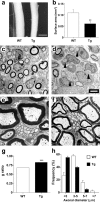
Figure 2.
Sciatic nerves of CNP-DN-erbB4 mice are thinner and show alterations in myelin and axonal morphology. a, Photomicrograph of freshly dissected sciatic nerves of 4-week-old WT and Tg mice. Scale bar, 370 μm. b, Cross-section surface area of sciatic nerve from 4-week-old transgenic was 45% thinner than that of wild-type mice (**p < 0.01; n = 3). c, d, Electron photomicrographs of sciatic nerve cross-sections of P7 wild-type (c) and CNP-DN-erbB4 (d) mice show that, in wild type, most large axons have segregated to form a 1:1 relationship with a Schwann cell and most large-caliber axons have formed significant myelin sheaths (c). In contrast, in the nerves of transgenic mice, most large-caliber axons have not formed myelin sheath (arrowheads), although they have been sorted and associated with Schwann cells (d). Scale bar, 2 μm. e, f, Electron microphotograph of sciatic nerve cross-sections of P28 wild-type (e) and transgenic (f) show that myelin is thinner in the mutant. Scale bar, 1 μm. g, g ratio in sciatic nerve fibers of P28 transgenic mice is larger than in wild type (***p < 0.001; n = 3). h, Distribution of axonal caliber in sciatic nerves. The proportion of small-diameter axons (<3 μm) is increased, whereas that of large-diameter axons (>5 μm) is decreased in transgenic mice (p < 0.001, χ2 test).
Figure 3.
Figure 3.
Reduction of internode length and nerve conduction velocity in CNP-DN-erbB4 mice. a, Microphotograph of an osmium-stained single sciatic nerve fiber of a 4-week-old mouse. The nodes of Ranvier are indicated by arrows and shown at higher magnification on the left. b, Internode length in transgenic mice is shorter than in wild type (***p < 0.001; n = 3). c, Tail nerve conduction velocity in 6-week-old transgenic mice is slower than in wild types (**p < 0.01; n = 9).
Figure 4.
Figure 4.
Transgenic mice have hypersensitivity to mechanical stimuli. a, Six-week-old mice were tested for mechanosensitivity using calibrated von Frey filaments. Transgenic mice show lower response threshold than wild-type mice. b, Transgenic and wild-type mice do not differ in their sensitivity to noxious thermal stimuli as measured by the hot-plate test.
Figure 5.
Figure 5.
Reduction in the level of expression of myelin genes in sciatic nerves of transgenic mice. a, The levels of P0, PMP22, and MBP mRNA in sciatic nerves of 4-week-old wild-type and DN-erbB4 mice were measured by real-time quantitative RT-PCR using 18S rRNA as a normalizer. Expression of the three genes was reduced in transgenic mice (*p < 0.05; ** p < 0.01; n = 3). b, Semiquantitative Western blot analysis of P0, PMP22, and MBP expression, using GAPDH as a normalizer, shows that the levels of these proteins are reduced in transgenic mice (*p < 0.05; ** p < 0.01; n = 3).
Figure 6.
Figure 6.
NRG1 isoform-specific effects on P0 expression in Schwann cells in vitro. a, Schwann cells in culture were either left untreated or were exposed to 1 nm EGF-like domain of NRG1 for 48 h, and the levels of P0 mRNA (left) or the activity of a P0-luciferase reporter construct (right) were measured. Both the mRNA levels and the activity of P0 promoter were increased by treatment with the EGF-like domain of NRG1 (**p < 0.01; n = 9). b, Top, Western blot analysis of HEK-293 cell lines stably expressing either type I or type III NRG1 with antibodies against the intracellular domain of NRG1 shows bands of the expected molecular weights. Bottom, Phosphotyrosine Western blot analysis of Schwann cells after coculture with the NRG1 HEK-293 cell lines for 20 min shows that both NRG1-expressing lines induce erbB receptor phosphorylation (p185) in Schwann cells, whereas the control cells do not. c, P0 mRNA in Schwann cells is increased fourfold by coculture with HEK-293 cells expressing type III NRG1, but contact with cells expressing type I NRG1 has no effect (*p < 0.05; n = 6).

References

    1. Arroyo EJ, Scherer SS (2000). On the molecular architecture of myelinated fibers. Histochem Cell Biol 113:1–18. - PubMed
    1. Black JA, Liu S, Tanaka M, Cummins TR, Waxman SG (2004). Changes in the expression of tetrodotoxin-sensitive sodium channels within dorsal root ganglia neurons in inflammatory pain. Pain 108:237–247. - PubMed
    1. Buonanno A, Fischbach GD (2001). Neuregulin and ErbB receptor signaling pathways in the nervous system. Curr Opin Neurobiol 11:287–296. - PubMed
    1. Carter GT, Jensen MP, Galer BS, Kraft GH, Crabtree LD, Beardsley RM, Abresch RT, Bird TD (1998). Neuropathic pain in Charcot-Marie-Tooth disease. Arch Phys Med Rehabil 79:1560–1564. - PubMed
    1. Chandross KJ, Cohen RI, Paras P Jr, Gravel M, Braun PE, Hudson LD (1999). Identification and characterization of early glial progenitors using a transgenic selection strategy. J Neurosci 19:759–774. - PMC - PubMed

Publication types

MeSH terms