. 2006 Mar 22;26(12):3220-8.
doi: 10.1523/JNEUROSCI.3780-05.2006.

Activation of kainate receptors sensitizes oligodendrocytes to complement attack

Affiliations

Activation of kainate receptors sensitizes oligodendrocytes to complement attack

Elena Alberdi et al. J Neurosci. .

Abstract

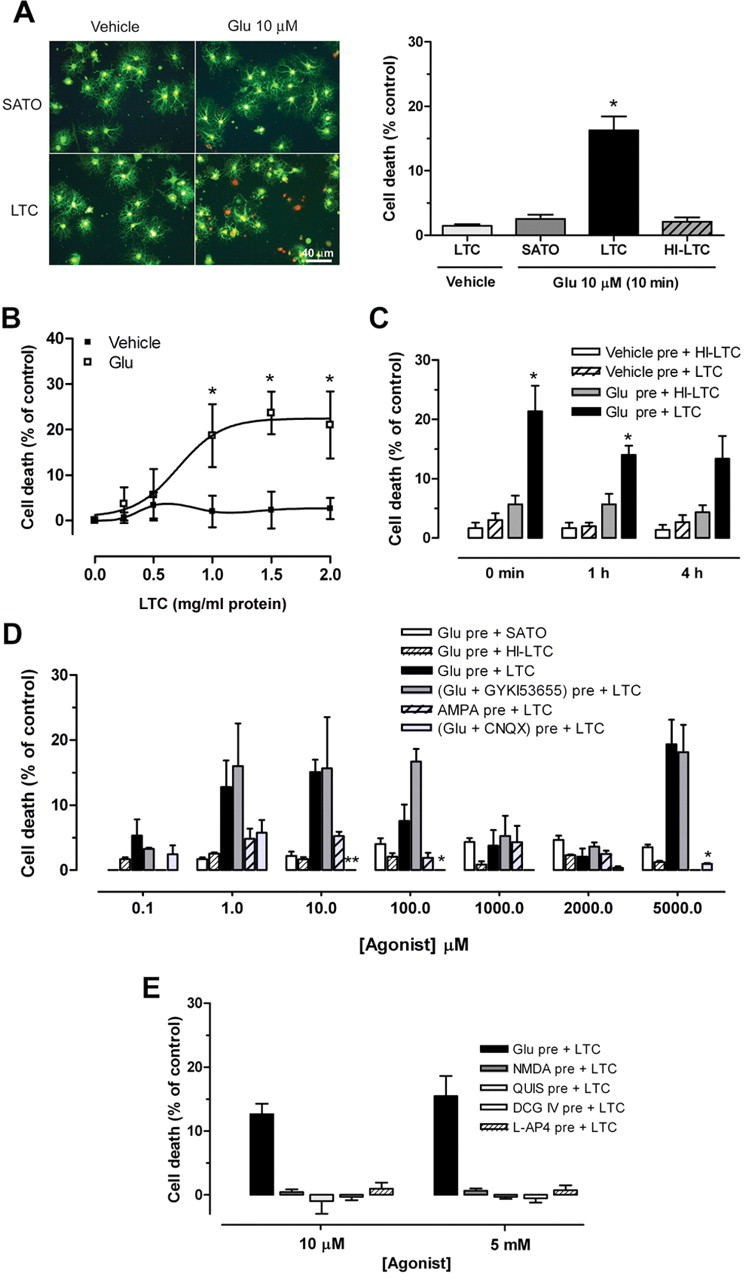
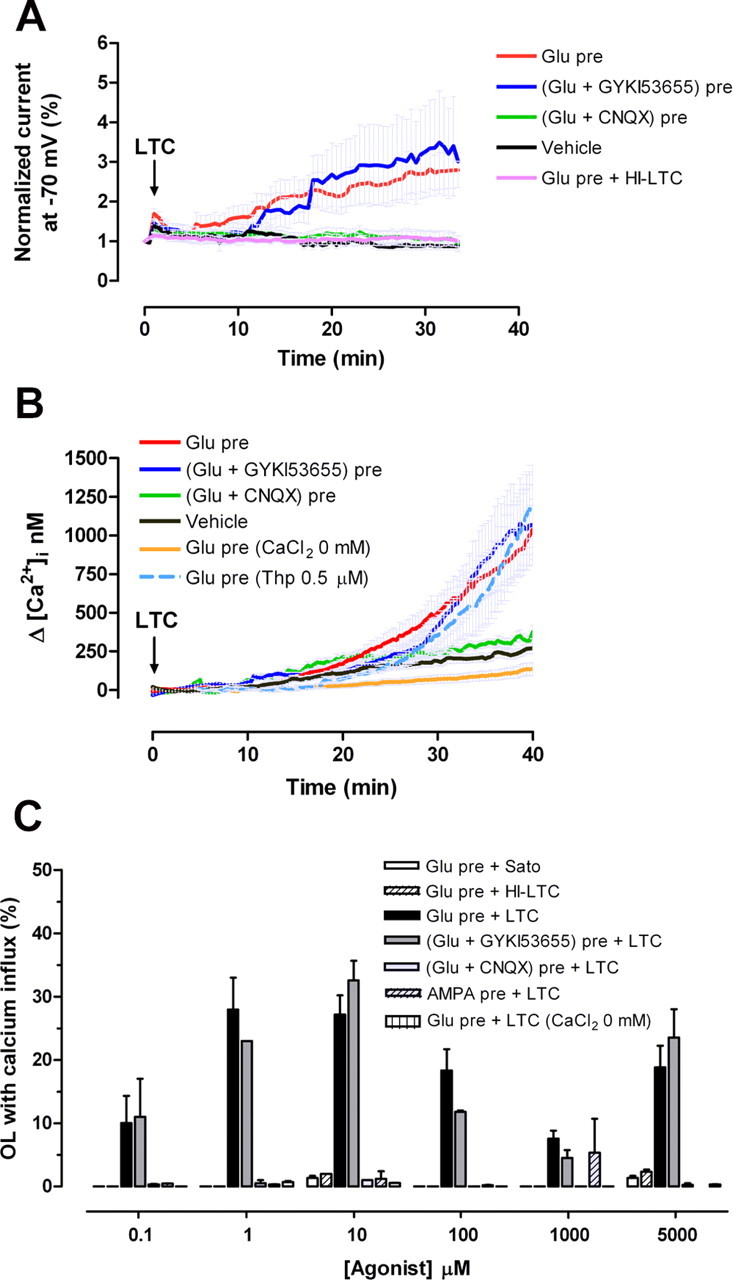
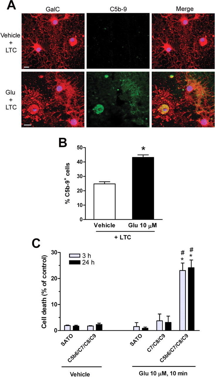
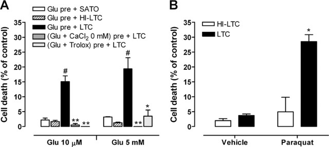
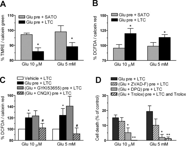
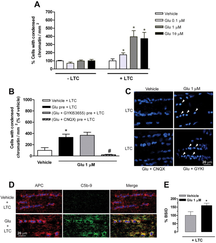
Glutamate excitotoxicity and complement attack have both been implicated separately in the generation of tissue damage in multiple sclerosis and in its animal model, experimental autoimmune encephalomyelitis. Here, we investigated whether glutamate receptor activation sensitizes oligodendrocytes to complement attack. We found that a brief incubation with glutamate followed by exposure to complement was lethal to oligodendrocytes in vitro and in freshly isolated optic nerves. Complement toxicity was induced by activation of kainate but not of AMPA receptors and was abolished by removing calcium from the medium during glutamate priming. Dose-response studies showed that sensitization to complement attack is induced by two distinct kainate receptor populations displaying high and low affinities for glutamate. Oligodendrocyte death by complement required the formation of the membrane attack complex, which in turn increased membrane conductance and induced calcium overload and mitochondrial depolarization as well as a rise in the level of reactive oxygen species. Treatment with the antioxidant Trolox and inhibition of poly(ADP-ribose) polymerase-1, but not of caspases, protected oligodendrocytes against damage induced by complement. These findings indicate that glutamate sensitization of oligodendrocytes to complement attack may contribute to white matter damage in acute and chronic neurological disorders.


Figures

Figure 1.
Figure 1.
Glutamate activation of kainate receptors sensitizes cultured oligodendrocytes to complement attack. A, Left, Oligodendrocyte viability (green calcein fluorescence) and death (propidium iodide red fluorescence) 24 h after pretreatment with vehicle or glutamate (Glu; 10 μm) for 10 min, followed by exposure to culture medium (SATO) or complement (LTC). Scale bar, 40 μm. Right, Histogram illustrating oligodendrocyte death. Note that HI-LTC was ineffective. Values are the mean ± SEM of duplicates from at least three different experiments.*p < 0.01, paired Student's t test. B, Dose–response curves showing LTC toxicity (0.25–2.00 mg/ml; 24 h) after pretreatment with vehicle or glutamate (10 μm, 10 min). Values are the mean ± SEM of duplicates from at least three different experiments. *p < 0.05 compared with vehicle pretreatment plus LTC, non-paired Student's t test. C, Time course of glutamate-mediated sensitization to complement attack. Cells were pretreated with vehicle or glutamate (10 μm, 10 min) and then incubated with LTC or HI-LTC immediately (0 min), 1 h, or 4 h later. Values are the mean ± SEM of duplicates from at least three different experiments. *p < 0.05 compared with vehicle pre + LTC or Glu pre + HI-LTC, paired Student's t test. D, Dose-dependent cell death after exposure to glutamate or AMPA as above. Cell death is maximal at 10 μm and 5 mm glutamate and is prevented by the AMPA/kainate antagonist CNQX (30 μm) but not by the AMPA-selective antagonist GYKI53655 (100 μm). In contrast, AMPA pretreatment induces very low toxicity. LTC alone (1.2 mg/ml) produced no detectable cell death. *p < 0.05, **p < 0.01 compared with Glu pre + LTC, paired Student's t test. E, NMDA and agonists of group I, II, and III metabotropic glutamate receptors quisqualate (QUIS), DCG IV, and l-AP-4, respectively, all at low and high concentrations, do not sensitize oligodendrocytes to complement toxicity. Values are the mean ± SEM of duplicates from at least three experiments.
Figure 2.
Figure 2.
Complement increases plasma membrane conductance and [Ca2+]i in glutamate-sensitized oligodendrocytes. A, Effect of complement (LTC) on oligodendroglial membrane conductance after pretreatment with glutamate (Glu; 5 mm for 10 min; n = 15). Current induced by LTC after glutamate pretreatment was blocked in the presence of CNQX (30 μm; n = 6) but not in the presence of GYKI53655 (100 μm; n = 8). Note that the membrane conductance of oligodendrocytes exposed exclusively to LTC (vehicle; n = 7) or to HI-LTC (n = 6) was not altered. B, Increase in [Ca2+]i in oligodendrocytes exposed to glutamate (Glu; 10 μm) and then to LTC is blocked by CNQX (30 μm) and by removal of Ca2+ from the medium but not by GYKI53655 (100 μm) or by Thp (0.5 μm). The curves illustrate average ± SEM responses of 15–52 cells from at least five different experiments. C, Percentage of oligodendrocytes showing massive Ca2+ influx during LTC incubation subsequent to exposure to different concentrations of glutamate or AMPA. Values represent average ± SEM of at least five experiments (n = 750 cells).
Figure 3.
Figure 3.
Glutamate pretreatment induces MAC formation in oligodendrocytes in culture. A, Cells were pretreated with vehicle (top) or glutamate (Glu; bottom; 10 μm, 10 min) and subsequently incubated with rabbit complement (LTC; 30 min). Cells are stained with antibodies to oligodendrocyte-type marker GalC (red; left) and to C5b-9 (green; middle) immunoreactivity. Colocalization of GalC and C5b-9 immunofluorescence is shown in merged images (yellow; right). Pretreatment with glutamate specifically induces MAC formation in oligodendrocyte. Blue fluorescence denotes oligodendrocyte nuclei as revealed by Hoechst 33258. Scale bar, 10 μm. B, Histogram represents the average ± SEM of double-labeled cells from duplicates of three different experiments. *p < 0.0001 compared with vehicle pretreatment plus LTC, non-paired Student's t test. C, Cell death at 3 and 24 h after pretreatment with vehicle or glutamate (10 μm, 10 min) followed by addition of MAC proteins C5b-6, C7, C8, and C9 (0.5 μg/ml each). Values represent average ± SEM. *p < 0.005 compared with vehicle pretreatment followed by C5b-6 plus C7/C8/C9; #p < 0.01 compared with glutamate pretreatment followed by C7/C8/C9.
Figure 4.
Figure 4.
Glutamate sensitization to complement attack in oligodendrocytes requires Ca2+ influx and the generation of ROS. A, Toxicity induced by complement (LTC) is prevented if glutamate (Glu) is added to the medium in the absence of Ca2+ or in the presence of the antioxidant Trolox (10 μm). Values represent the mean ± SEM of duplicates from three different experiments. #p < 0.005 compared with Glu pre + SATO; *p < 0.05, **p < 0.01 compared with Glu pre + LTC; paired Student's t test. B, Paraquat-sensitized oligodendrocytes to complement attack. Cells were exposed to vehicle or 100 μm paraquat for 3 h and then exposed to HI-LTC or LTC for an additional 24 h. *p < 0.005 compared with vehicle pretreatment followed by LTC, paired Student's t test.
Figure 5.
Figure 5.
Complement causes mitochondrial depolarization (A), oxidative stress (B, C), and PARP-1-mediated apoptosis in glutamate (Glu)-sensitized oligodendrocytes (D). Each culture measurement was normalized to calcein fluorescence (A) or calcein red–orange fluorescence (B, C), indicators of cell viability, and 100% represents control values in the absence of agonist. D, PARP-1 inhibition with DPQ (30 μm) reduces cell death, whereas the pan-caspase inhibitor ZVAD-F (50 μm) was ineffective. In turn, the antioxidant Trolox (10 μm) fully protected oligodendrocytes from complement (LTC) toxicity. Values represent mean ± SEM of triplicates from six different experiments. A, B, *p < 0.05 compared with Glu pre + SATO. C, *p < 0.05 compared with vehicle + LTC; #p < 0.05 compared with Glu pre + LTC. D, *p < 0.05, **p < 0.01 compared with Glu pre + LTC. All by paired Student's t test.
Figure 6.
Figure 6.
Glutamate sensitizes oligodendrocytes in situ to complement attack. A, Maximal sensitization to complement (LTC) attack is observed after pretreatment of isolated optic nerves with 1 μm glutamate (Glu). Values (average ± SEM) were obtained from at least three optic nerves. *p < 0.01 by non-paired Student's t test. B, Glutamate with CNQX, but not with GYKI536555, significantly reduces the number of cells with condensed chromatin in optic nerves incubated with LTC. Values (average ± SEM) were obtained from at least three optic nerves. *p < 0.05 compared with vehicle + LTC; #p < 0.001 compared with Glu pre + LTC. C, Representative images showing nuclei of optic nerve cells labeled with Hoechst 33258 after exposure to artificial CSF (vehicle) and glutamate alone or in conjunction with CNQX and GYKI53655, followed by LTC. Notice that cells with condensed chromatin (arrows) are only evident in conditions in which kainate receptors are activated by glutamate. D, Formation of MAC (green) around the oligodendrocytes (red) is absent in optic nerves treated with vehicle (top row) but occurs in nerves incubated with glutamate (bottom row), as visualized with anti-C5b-9 (green) and APC antibodies (red). Colocalization of APC and C5b-9 immunofluorescence is shown in merged images (yellow; right). Nuclei are labeled with Hoechst 33258. E, Quantification of MAC immunofluorescence in nerves treated with vehicle or glutamate followed by LTC. Values (average ± SEM) were obtained from four optic nerves and at least 20 fields per nerve were evaluated.*p < 0.05 by paired Student's t test. Scale bars, 20 μm.

References

    1. Alberdi E, Sánchez-Gómez MV, Marino A, Matute C (2002). Ca2+ influx through AMPA or kainate receptors alone is sufficient to initiate excitotoxicity in cultured oligodendrocytes. Neurobiol Dis 9:234–243. - PubMed
    1. Appel SH, Bonstein MB (1964). The application of tissue culture to the study of experimental allergic encephalomyelitis. II. Serum factors responsible for demyelination. J Exp Med 119:303–312. - PMC - PubMed
    1. Barnett MH, Prineas JW (2004). Relapsing and remitting multiple sclerosis: pathology of the newly forming lesion. Ann Neurol 55:458–468. - PubMed
    1. Barnum SR (1995). Complement biosynthesis in the central nervous system. Crit Rev Oral Biol Med 6:132–146. - PubMed
    1. Bohana-Kashtan O, Ziporen L, Donin N, Kraus S, Fishelson Z (2004). Cell signals transduced by complement. Mol Immunol 41:583–597. - PubMed

Publication types

MeSH terms

LinkOut - more resources