. 2006 Mar 22;26(12):3256-68.
doi: 10.1523/JNEUROSCI.5537-05.2006.

Motoneurons derived from embryonic stem cells express transcription factors and develop phenotypes characteristic of medial motor column neurons

Affiliations

Motoneurons derived from embryonic stem cells express transcription factors and develop phenotypes characteristic of medial motor column neurons

Prabakaran Soundararajan et al. J Neurosci. .

Abstract

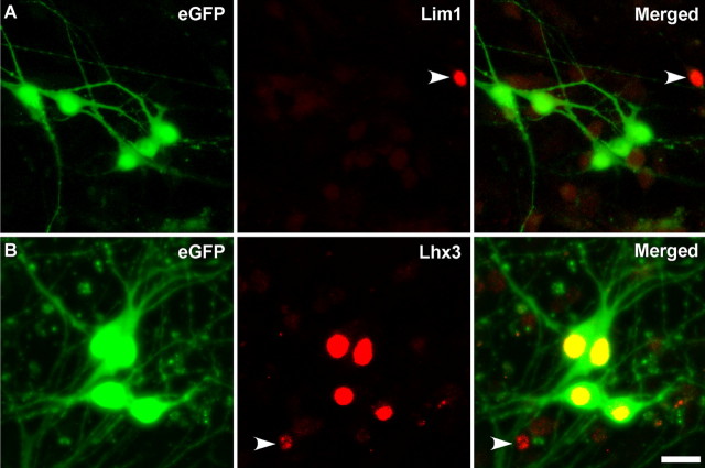
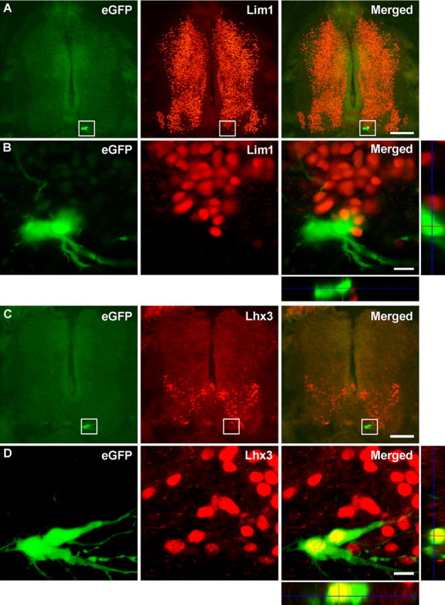
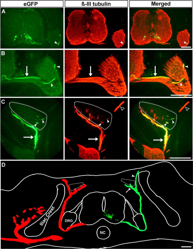
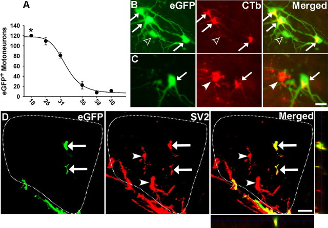
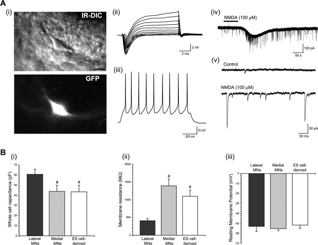
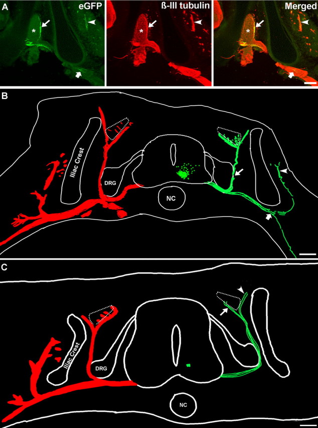
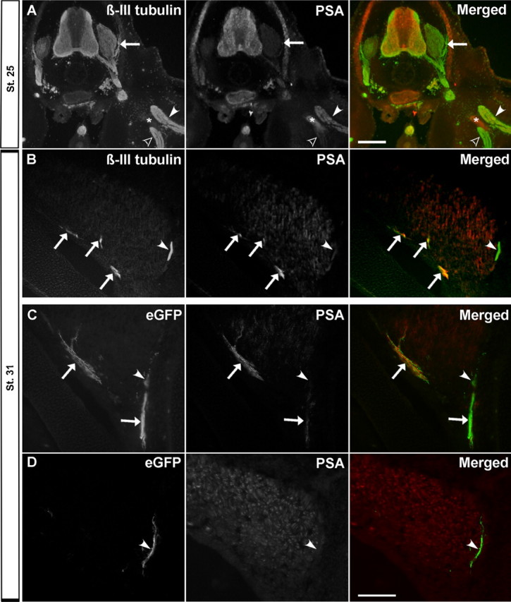
Embryonic stem (ES) cells differentiate into functional motoneurons when treated with a sonic hedgehog (Shh) agonist and retinoic acid (RA). Whether ES cells can be directed to differentiate into specific subtypes of motoneurons is unknown. We treated embryoid bodies generated from HBG3 ES cells with a Shh agonist and RA for 5 d in culture to induce motoneuron differentiation. Enhanced green fluorescent protein (eGFP) expression was used to identify putative motoneurons, because eGFP is expressed under the control of the Hb9 promoter in HBG3 cells. We found that 96 +/- 0.7% of the differentiated eGFP+ motoneurons expressed Lhx3, a homeobox gene expressed by postmitotic motoneurons in the medial motor column (MMCm), when the treated cells were plated on a neurite-promoting substrate for 5 d. When the treated embryoid bodies were transplanted into stage 17 chick neural tubes, the eGFP+ motoneurons migrated to the MMCm, expressed Lhx3, projected axons to the appropriate target for MMCm motoneurons (i.e., epaxial muscles), and contained synaptic vesicles within intramuscular axonal branches. In ovo and in vitro studies indicated that chemotropic factors emanating from the epaxial muscle and/or surrounding mesenchyme likely guide Lhx3+ motoneurons to their correct target. Finally, whole-cell patch-clamp recordings of transplanted ES cell-derived motoneurons demonstrated that they received synaptic input, elicited repetitive trains of action potentials, and developed passive membrane properties that were similar to host MMCm motoneurons. These results indicate that ES cells can be directed to form subtypes of neurons with specific phenotypic properties.


Figures

Figure 1.
Figure 1.
ES cell-derived motoneurons continually express Lhx3 in culture when previously exposed to RA and Hh-Ag1.3. A, B, The majority of eGFP+ cells had extensive neurites 5 d after plating. A, Immunolabeling showed that very few of the eGFP+ ES cell-derived motoneurons expressed Lim1, although eGFP/Lim1+ cells were occasionally observed (A, arrowhead). B, In contrast, the vast majority of the eGFP+ cells, and a few eGFP cells (B, arrowhead), expressed Lhx3. Scale bar, 20 μm.
Figure 2.
Figure 2.
ES cell-derived motoneurons translocate to the MMCm and continue to express Lhx3 when transplanted in ovo. A, C, Cross sections through st. 31 chick embryos showed that the majority of the transplanted eGFP+ cells translocated to the medial/ventral aspect of the developing neural tube. A, B, Immunolabeling and confocal imaging showed that none of the transplanted eGFP+ cells were Lim1+. B, Images are a higher magnification of the corresponding boxes shown in A. Imaging in both the xz and yz orthogonal planes confirm that the eGFP+ cells were Lim1. C, D, Although less intensely stained than the endogenous chick Lhx3+ neurons, all of the transplanted eGFP+ cells were Lhx3+. D, Images are a higher magnification of the boxes shown in C. Imaging in both the xz and yz orthogonal planes confirms the expression of Lhx3 in eGFP+ cells (D). In all sections, dorsal is up. Scale bars: A, C, 250 μm; B, D, 20 μm.
Figure 3.
Figure 3.
Transplanted ES cell-derived motoneurons project axons to axial muscles. A, D, Cross sections through st. 31 chick embryos showed that the majority of the transplanted eGFP+ cells translocated to the medial/ventral aspect of the neural tube. B, ES cell-derived motoneurons extended axons out of the neural tube through the ventral root (B, arrow) and projected dorsally around the DRG as part of the dorsal ramus (A, B, arrowheads). C, Dorsal projecting eGFP+ axons formed part of the dorsal ramus (C, arrow) and innervated the longissimus muscle (C, arrowheads; muscle delineated by dotted line). The endogenous cutaneous nerve is indicated with an open arrowhead. A–C, β-III tubulin immunohistochemistry was used to visualize both the endogenous chick and transplanted mouse neurons. D, Neurolucida reconstruction shows that all of the eGFP+ axons in a representative st. 31 chick embryo (green tracings) projected dorsally and either formed part of the longissimus muscle nerve (D, arrow) or incorrectly targeted the skin (D, arrowhead). Each green dot in the neural tube represents one eGFP+ neuron. For comparison, the projection patterns of the endogenous β-III tubulin+ chick neurons are shown in red on the contralateral side. The longissimus muscles are indicated with dotted lines. In all sections, dorsal is up. NC, Notochord. Scale bars: A, D, 100 μm; B, C, 200 μm.
Figure 4.
Figure 4.
Transplanted ES cell-derived motoneurons survive beyond the cell death period, innervate the epaxial muscle, and express presynaptic proteins required for synapse formation. A, The number of eGFP+ cells in the neural tube was quantified before (st. 25), during (st. 31 and 36), and after (st. 38 and 40) the naturally occurring cell death period to characterize the degree and pattern of cell survival. The asterisk denotes that the cell number was an estimate. The curve was fitted according to the formula: y = min + (max − min)/(1 + (x/EC50)Hillslope) and had an r2 value of 0.998. Error bars indicate SE. B, C, Cross sections through the MMCm shows eGFP+ cell bodies that either contained (arrows) or did not contain (open arrowhead) the retrograde tracer CTb. A backlabeled endogenous MMCm neuron is also present (C, arrowhead). D, Confocal imaging shows that the eGFP+ axons innervating the longissimus muscle (outlined by dotted line) were immunolabeled with a synaptic vesicle-specific antibody (SV2; arrows). Endogenous eGFP chick neurons are indicated with arrowheads. Imaging in both the xz and yz orthogonal planes confirm that the eGFP+ axons were SV2+. Scale bars: (in C) B, C, 30 μm; D, 20 μm.
Figure 5.
Figure 5.
Transplanted ES cell-derived motoneurons express guidance molecules appropriate for MMCm motoneurons. A, Cross section shows that the axon shaft (arrow), but not the distal tips (open arrowhead), of β-III tubulin+ neurons projecting along the ventral edge of the longissimus muscle expressed EphA4 in control st. 31 chick embryos. As expected, the cutaneous axons projecting to the skin were EphA4 (arrowhead). B, Confocal imaging shows that the eGFP+ axons from the transplanted ES cell-derived motoneurons expressed EphA4 as they correctly projected along the ventral border of the longissimus muscle (arrow) and incorrectly projected to the skin (arrowhead). Imaging in the xz orthogonal plane confirms that the eGFP+ axons were EphA4+. Scale bar, 100 μm.
Figure 6.
Figure 6.
Chemotropic mechanisms selectively guide ES cell-derived motor axons to their correct target. A, Cartoon showing the location of the Hh-Ag1.3-treated embryoid bodies that were deliberately transplanted into the dorsal hemisphere of st. 17 chick neural tubes. B, Neurolucida reconstruction shows that the transplanted ES cell-derived motoneurons were primarily located in the dorsal one-half of the neural tube in a st. 31 chick embryo (green dots). The eGFP+ axons (green tracings) extended out of the neural tube along the dorsal root (long arrow) where they later projected dorsally as part of the dorsal ramus. The dorsal projecting eGFP+ axons later bifurcated to either form part of the longissimus muscle nerve (short arrow) or cutaneous nerve that innervates the skin. C, Higher magnification of the box shown in B immunolabeled with β-III tubulin (red). eGFP+/β-III tubulin+ axons appear yellow in this merged image. Arrowhead shows an eGFP+ axon extending away from the endogenous chick dorsal root axons. D, A z-stack collapsed confocal image of an Hh-Ag1.3/RA-treated embryoid body that was flanked with explants harvested from the longissimus and caudilioflexorius muscles showing that the neurite outgrowth was substantially greater on the side of the longissimus muscle. The picture is shown as an inverted black and white image of the green fluorescence. Scale bars, 100 μm. E, Quantitative analysis indicates that significantly more neurites grew toward the longissimus muscle explant compared with the caudilioflexorius explant (n = 8; *p = 0.0004). Error bars indicate SE.
Figure 7.
Figure 7.
ES cell-derived motoneurons transplanted into the developing chick spinal cord express appropriate electrophysiological properties. Ai, An ES cell-derived motoneuron in a chick spinal cord slice visualized using IR-DIC microscopy (top) and epifluorescence exciting GFP (bottom). Scale bar, 10 μm. Aii, A voltage-clamp recording from an ES cell-derived motoneuron showing fast, inactivating inward currents and persistent outward currents in response to depolarizing voltage steps from a holding potential of −60 mV. Aiii, A current-clamp recording from an ES cell-derived motoneuron showing repetitive firing of action potentials in response to the injection of a 1 s current pulse. Aiv, A voltage-clamp recording of an ES cell-derived motoneuron held at −60 mV showing an inward current in response to the application of NMDA (100 μm). Av, Recordings in Aiv shown on a faster time scale demonstrating the presence of EPSCs in control conditions and during the application of NMDA. Bi–Biii, Graphs comparing the passive membrane properties of endogenous lateral and medial motoneurons (MNs) and transplanted ES cell-derived motoneurons. The pound sign indicates significant difference compared with lateral motoneuron. Error bars indicate SE.
Figure 8.
Figure 8.
Transplanted ES cell-derived motoneurons can project axons to limb muscles. A, Cross sections through a st. 31 chick embryo transplanted with ∼275 ES cell-derived motoneurons cells shows that the eGFP+ axons either projected dorsally along the dorsal ramus (A, B, long arrow), or extended along other major peripheral axonal pathways that ultimately led to the ilio-femoralis internus muscle (A, B, arrowhead) or into the developing limb (A, B, short arrow). B, Neurolucida reconstruction shows all of the eGFP+ motoneurons (green tracings) in a representative st. 31 chick embryo transplanted with ∼275 ES cell-derived motoneurons. For comparison, the projection patterns of the endogenous β-III tubulin+ chick neurons are shown in red on the contralateral side. C, Neurolucida reconstruction shows that some eGFP+ motor axons misproject to the skin even when only 10 eGFP+ cells are transplanted into the neural tube. NC, Notochord. Scale bars: A, 20 μm; B, C, 100 μm.
Figure 9.
Figure 9.
ES cell-derived motor axons projecting to the epaxial muscle and skin differentially express PSA. A, Cross section of a control st. 25 embryo immunostained for β-III tubulin and PSA shows that PSA is not expressed by axons as they form the dorsal ramus (arrow). As expected, the dorsal nerve trunk projecting into the hindlimb expressed more PSA (arrowhead) than the ventral nerve truck (open arrowhead). The asterisk denotes autofluorescing bone. B, At stage 31, the muscle component of the dorsal ramus expresses PSA (arrows), whereas the sensory component does not (arrowhead). C, D, eGFP+ axons projecting to the longissimus muscle express high levels of PSA (arrow), whereas the eGFP+ axons extending to the skin along the cutaneous nerve do not (arrowheads). In all sections, dorsal is up. Scale bars: A, 100 μm; (in D) B–D, 200 μm.

Similar articles

Cited by

References

    1. Abercrombie M (1946). Estimation of nuclear population from microtome sections. Anat Neurobiol Rec 94:239–247. - PubMed
    1. Bjorklund LM, Isacson O (2002). Regulation of dopamine cell type and transmitter function in fetal and stem cell transplantation for Parkinson's disease. Prog Brain Res 138:411–420. - PubMed
    1. Chisholm A, Tessier-Lavigne M (1999). Conservation and divergence of axon guidance mechanisms. Curr Opin Neurobiol 9:603–615. - PubMed
    1. Dahm LM, Landmesser LT (1991). The regulation of synaptogenesis during normal development and following activity blockade. J Neurosci 11:238–255. - PMC - PubMed
    1. Dasen JS, Tice BC, Brenner-Morton S, Jessell TM (2005). A hox regulatory network establishes motor neuron pool identity and target-muscle connectivity. Cell 123:477–491. - PubMed

Publication types

MeSH terms