. 2006 Mar 22;26(12):3319-29.
doi: 10.1523/JNEUROSCI.5383-05.2006.

Axonal ensheathment and septate junction formation in the peripheral nervous system of Drosophila

Affiliations

Axonal ensheathment and septate junction formation in the peripheral nervous system of Drosophila

Swati Banerjee et al. J Neurosci. .

Abstract

Axonal insulation is critical for efficient action potential propagation and normal functioning of the nervous system. In Drosophila, the underlying basis of nerve ensheathment is the axonal insulation by glial cells and the establishment of septate junctions (SJs) between glial cell membranes. However, the details of the cellular and molecular mechanisms underlying axonal insulation and SJ formation are still obscure. Here, we report the characterization of axonal insulation in the Drosophila peripheral nervous system (PNS). Targeted expression of tau-green fluorescent protein in the glial cells and ultrastructural analysis of the peripheral nerves allowed us to visualize the glial ensheathment of axons. We show that individual or a group of axons are ensheathed by inner glial processes, which in turn are ensheathed by the outer perineurial glial cells. SJs are formed between the inner and outer glial membranes. We also show that Neurexin IV, Contactin, and Neuroglian are coexpressed in the peripheral glial membranes and that these proteins exist as a complex in the Drosophila nervous system. Mutations in neurexin IV, contactin, and neuroglian result in the disruption of blood-nerve barrier function in the PNS, and ultrastructural analyses of the mutant embryonic peripheral nerves show loss of glial SJs. Interestingly, the murine homologs of Neurexin IV, Contactin, and Neuroglian are expressed at the paranodal SJs and play a key role in axon-glial interactions of myelinated axons. Together, our data suggest that the molecular machinery underlying axonal insulation and axon-glial interactions may be conserved across species.


Figures

Figure 1.
Figure 1.
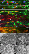
Glial cell profile in the embryonic and larval PNS. A–C, Stage 13 wild-type (A) and repo-GAL4::UAS-tauGFP (B, C) embryos stained with anti-Repo (green; A) and anti-Fas II (red; A–C) show that the clusters of peripheral glial cells begin to migrate to the periphery along the motor axon tracts. The anti-GFP (green; B, C) shows the profile of the glial microtubular cytoskeleton, thus highlighting the glial process extension. C, Higher magnification of a portion of the embryo shown in B reveals that the glial cytoplasmic processes (arrow) extend along the motor axon tracts (arrowhead) to reach the periphery. The CNS–PNS boundary is highlighted by a white line (C, F, I). D–F, Stage 14 wild-type (D) and repo-GAL4::UAS-tauGFP (E, F) embryos show Repo-positive glial cells (green; D) moving further into the periphery along the motor axons (red; D). The tau-GFP visualized by anti-GFP staining (green; E, F) shows the extent of the glial microtubular network (arrow; F) along the axonal tracts (arrowhead; F). The group of lateral chordotonal sensory organs (lch) is also visible in this focal plane and shows expression of GFP in ligament cells, which ensheath chordotonal neurons. G–I, Stage 16 wild-type (G) and repo-GAL4::UAS-tauGFP (H, I) embryos show that the Repo-positive glial cells (green; G) have migrated to the periphery, and the glial processes (green; H, I) visualized by anti-GFP staining show near complete ensheathment of the peripheral nerve (arrow; I) axon tracts (red; H, I). The glial ensheathment has extended up to lch. The axon tracts are seen running through the glial cells. The sensory bipolar dendritic neuron is also ensheathed by its supporting glial cell (green arrow). J, A portion of the third-instar larval ventral nerve cord with peripheral nerves expressing GFP under repo-GAL4 driver. The glial nuclei (arrows) are randomly lined up along the peripheral nerves, and no consistent distance is observed between neighboring glial cells. K, A portion of an abdominal nerve with a glial cell nucleus (N) from repo-GAL4::UAS-tauGFP stained with anti-GFP reveals the pattern of the glial microtubular network (arrows) with tracts of microtubules separated from each other. L–Q, A portion of a peripheral nerve of repo-GAL4::UAS-tauGFP stained with anti-GFP (L, O) and anti-Fas II (M, P) reveals the ensheathment of the nerve by the glial membranes (N, Q). The glial membrane extends partly into the initial portion of the third-instar larval neuromuscular junctions (arrows; N, Q) (Sepp et al., 2000). The glial processes also display arborizations at the initial contact site with the muscle (Q, arrows). The Fas II marked synaptic boutons (arrowheads; N, Q) that are away from the nerve entry site are devoid of glial insulation and are protected by the musculature.
Figure 2.
Figure 2.
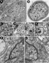
Axonal ensheathment and presence of junctions in the nerve fibers. A, Cross section through a 20- to 22-h-old wild-type embryonic nerve fiber showing tightly held axons (a) surrounded by a glial cell (g). The electron-dense region where two glial membranes come in close apposition is highlighted by an arrow. B, Cross section through a wild-type third-instar peripheral nerve at a low magnification shows the arrangement of axons (a) surrounded by glial membranes (m). The outer perineurial glial cell (P) is surrounding the axons (a) and the inner glial membranes (m). The nerve is surrounded by a neural lamella (l). Note the presence of electron-dense areas that contain SJs (arrow). C, Section through a peripheral nerve showing glial membrane (m) interspersed between the axons (a). D, A nerve fascicle containing a group of axons is collectively wrapped by the inner glial membrane (m). The arrowheads point to the boundary of the glial membrane that ensheaths this fascicle. Some axons (a) in the peripheral nerves are individually wrapped by the inner glial membrane (E, arrowheads). F, A portion of the same nerve as in B at a higher magnification shows axons (a) surrounded by glial membrane processes (m). The inner and outer glial membranes come in close proximity to form the SJs (arrows). Glial membranes with SJs always display a distinct electron dense ladder-like structure corresponding to glial–glial SJs (F, G, arrows). Scale bars: A, D, 0.5 μm; B, C, 1 μm; E, 0.25 μm; F, G, 0.1 μm.
Figure 3.
Figure 3.
Localization of Nrx IV, Cont, and Nrg in the embryonic nervous system. A, A portion of a wild-type stage 16 embryo stained for Nrx IV (green), Fas II (red), and Repo (blue). Nrx IV localizes to the glial membrane (arrowhead), whereas Fas II stains the motor axons (arrow). The glial nuclei are lined up along the length of the nerve fiber (blue). Cont (B; green, arrowhead) and Nrg (C; green, arrowhead) also localize to the glial membrane. However, Nrg has additional localization in the neurons (C; yellow, arrow). D–F, A segment of a stage 16 gcm mutant embryo shows an absence of Repo-positive glial cells (blue) and a lack of glial membrane localization of Nrx (green; D), Cont (green; E), and Nrg (green, F). However, the epithelial localization of Nrx and Cont (D, E, respectively) as well as the neuronal localization of Nrg (F) in gcm mutant embryos remain unaltered (compare with wild type in A–C). Sensory and motor neurons are stained by anti-HRP (red; F). G, A cross section through a wild-type embryonic peripheral nerve fiber shows ensheathment of the axons by the glial membrane and close apposition of glial membranes where SJs are observed (black arrowhead). H, A cross section through a gcm mutant embryonic peripheral nerve shows the absence of a glial membrane (black arrowhead). However, the axons (a) are still held together as reported previously by Jones et al. (1995). I, A portion of the wild-type nerve from G at a higher magnification shows glial membranes (black arrows) in close apposition surrounding the axons (a). J, A portion of a gcm mutant peripheral nerve lacks glial membranes (black arrowheads) around the axons (a). Scale bars: G, H, 0.5 μm; I, J, 0.2 μm.
Figure 4.
Figure 4.
Interdependence and protein complex formation between Nrx, Cont, and Nrg in the nervous system. A portion of a wild-type stage 16 embryo stained for Nrx IV (A), Nrg (B), and Cont (C). Nrx IV, Cont, and Nrg colocalize in the glial membrane (A–D; arrows). E–H, A stage 16 nrx IV null embryo stained with anti-Nrx IV (E), anti-Cont (F), and anti-Nrg (G) shows highly diffused localization of Cont (F) in the glial membranes (compare with C). Cont is also localized extensively in small puncta in the cytoplasm of the glial cells (arrowheads; inset in H). Nrg (G) appears diffused along the nerve fibers (compare with B). I–L, A stage 16 cont null embryo stained with anti-Cont (I), anti-Nrx IV (J), and anti-Nrg (K) shows diffused localization of Nrx IV (J) and Nrg (K) (arrowheads; inset in L), compared with their respective wild-type localization in A and B, respectively. M–P, A stage 16 nrg null embryo stained with anti-Nrg (M), anti-Nrx IV (N), and anti-Cont (O) shows reduced levels of Nrx IV (N, compare with wild-type A) and Cont (O, compare with C) in the glial membrane (arrowheads; inset in P). Coimmunoprecipitation experiments reveal a tripartite complex between Nrx IV, Cont, and Nrg. Adult head lysates were immunoprecipitated with anti-Nrx IV, anti-Cont, and anti-Nrg antibodies, and the immunoprecipitated proteins were resolved by SDS-PAGE and immunoblotted with anti-Nrx IV (Q), anti-Cont (R), and anti-Nrg antibodies (S). All three proteins were immunoprecipitated by their cognate antibodies and also coprecipitated the other two proteins. All antibodies coprecipitated both isoforms of Nrg (167 and 180 kDa isoforms). The PIs were used as controls, which did not precipitate any of these proteins. T, Sucrose density gradient centrifugation of head lysates shows cosedimentation of Nrx IV and Cont and partial cosedimentation of Nrg with Nrx IV and Cont, suggesting that these proteins may exist as a biochemical complex in vivo. U, Immunoblots of the 20-h-old wild-type, nrx IV, cont, and nrg mutant embryos probed with antibodies against Nrx IV (Ua), Cont (Ub), and Nrg (Uc) show dramatic reduction of Cont and Nrg in nrx IV mutants. For protein level control, the blots were probed with anti-Barren, an unrelated protein (Ud) (Bhat et al., 1996).
Figure 5.
Figure 5.
Disruption in CO morphology and BNB function in nrx IV, cont, and nrg mutants. A, Schematic of a CO showing the various cell types. Cap cells (cc; green), scolopale (s.c.; red), and ligament (lig) are the three glial cell types. The neuron (neu; blue) has a rootlet (r), and its dendrite (d) projects into the lumen (lu) of the scolopale. The cc and the lig cell attachment sites of the CO to the epidermis (ep) are also shown. The presence of extensive SJs has been reported between the cap and scolopale cells, thus providing a functional BNB. B, Wild-type CO triple stained with anti-β3 tubulin (green) marking the cap cells, anti-Crb (red) marking the lumen of the scolopale, and anti-22C10 (blue) marking the sensory neuron. C–H, Wild-type COs (C, E, G) stained with anti-CRB (red) and anti-22C10 (blue) in combination with anti-Nrx IV (C, D), anti-Cont (E, F), and anti-Nrg (G, H) show a fusiform shape of the scolopales in the CO cluster. nrx IV (D), cont (F), and nrg (H) mutants as evident from the lack of staining of their respective antibodies show a defective morphology and a disarrayed organization of the cluster. I–L, Dye exclusion assays performed on the wild-type embryos (I), nrx IV (J), cont (K), and nrg (L) mutant embryos. Confocal images after dye injection of the regions of the peripheral nervous system at the level of the COs are shown. Wild-type embryos (I) excluded the dye from the COs even after 30 min of injection, indicating that a functional BNB is present. Under identical conditions nrx IV (J), cont (K), and nrg (L) mutant embryos failed to exclude the dye from the COs. Confocal images showed dye penetration into COs within 15 min after injection, indicating that the BNB has broken down in these mutants.
Figure 6.
Figure 6.
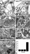
nrx IV, cont, and nrg null mutant embryos lack glial SJs. A, Ultrastructure of a portion of a wild-type embryonic nerve shows the presence of distinct ladder-like SJs between the glial membranes (arrows). Also note the smooth membrane surfaces of the axons and the inner glial membrane (arrowheads). B, Ultrastructure of a portion of a nrx IV null mutant embryo shows loss of SJs between the glial membranes (arrows) and a fuzzy appearance at the axon-glial interface (arrowheads). Axons are marked as a, and glial cells are marked as g. C, E, Electron micrograph of a cross section through a wild-type peripheral nerve fiber at increasing magnifications reveals the presence of extensive glial SJs (arrows) and axons (a). D, F, Electron micrograph through a cross section of nrg mutant peripheral nerve fiber at increasing magnifications show complete lack of glial SJs (arrows) and increased spacing between the glial membranes. Note the missing axons in the nrg mutant nerves (asterisks; D, F). G, Ultrastructure of a cross section of cont mutant peripheral nerve shows loss of glial SJs (arrows) without any change in the intermembrane spacing, a phenotype that is similar to that of nrx IV mutants. H, Statistical analysis of glial intermembrane spacing in wild-type, nrx IV, cont, and nrg mutant nerves. The intermembrane spacing did not seem significantly different between the wild-type, nrx IV, and cont mutants. The intermembrane spacing between the outer and inner glial membranes in nrg mutant showed a dramatic increase when compared with wild-type nerves. Scale bars: A, B, E–G, 0.2 μm; C, D, 0.5 μm.

References

    1. Anderson JM, Van Itallie CM, Fanning AS (2004). Setting up a selective barrier at the apical junction complex. Curr Opin Cell Biol 16:140–145. - PubMed
    1. Auld VJ, Fetter RD, Broadie K, Goodman CS (1995). Gliotactin, a novel transmembrane protein on peripheral glia, is required to form the blood-nerve barrier in Drosophila. Cell 81:757–767. - PubMed
    1. Bainton RJ, Tsai LT, Schwabe T, DeSalvo M, Gaul U, Heberlein U (2005). moody encodes two GPCRs that regulate cocaine behaviors and blood–brain barrier permeability in Drosophila. Cell 123:145–156. - PubMed
    1. Banerjee S, Sousa AD, Bhat MA (2006). Organization and function of septate junctions: an evolutionary perspective. Cell Biochem Biophys in press. - PubMed
    1. Baumgartner S, Littleton JT, Broadie K, Bhat MA, Harbecke R, Lengyel JA, Chiquet-Ehrismann R, Prokop A, Bellen HJ (1996). A Drosophila neurexin is required for septate junction and blood-nerve barrier formation and function. Cell 87:1059–1068. - PubMed

Publication types

MeSH terms

LinkOut - more resources