. 2006 Mar 22;26(12):3335-44.
doi: 10.1523/JNEUROSCI.5010-05.2006.

An analog of a dipeptide-like structure of FK506 increases glial cell line-derived neurotrophic factor expression through cAMP response element-binding protein activated by heat shock protein 90/Akt signaling pathway

Affiliations

An analog of a dipeptide-like structure of FK506 increases glial cell line-derived neurotrophic factor expression through cAMP response element-binding protein activated by heat shock protein 90/Akt signaling pathway

Xiaobo Cen et al. J Neurosci. .

Abstract

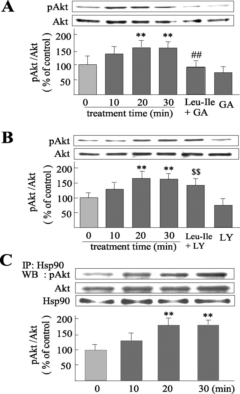
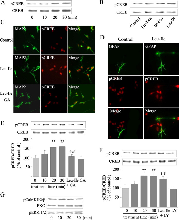
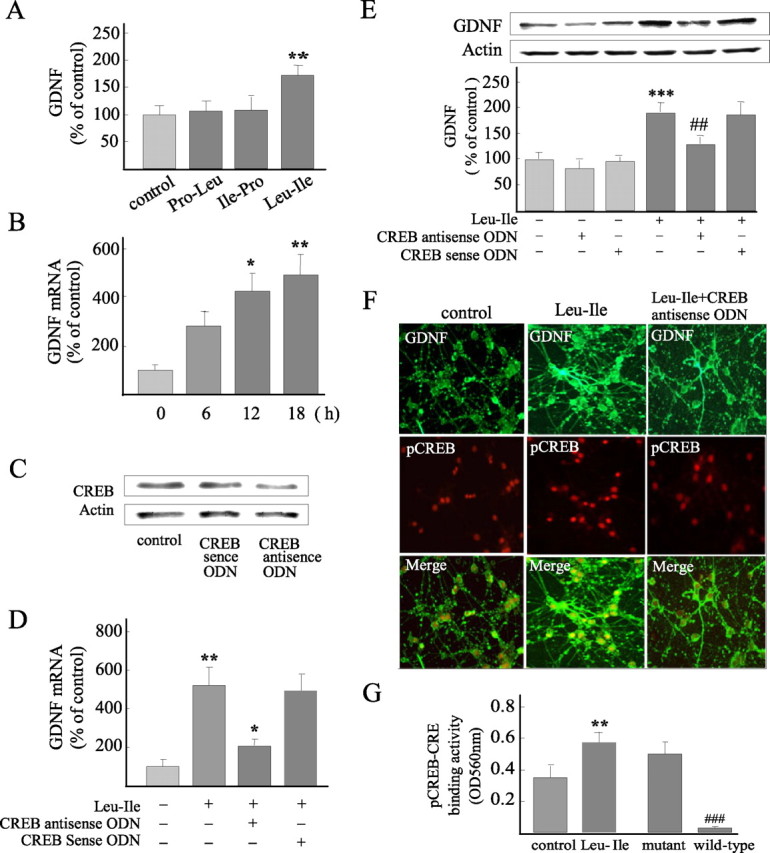
Glial cell line-derived neurotrophic factor (GDNF) is an important neurotrophic factor that has therapeutic implications for neurodegenerative disorders. We previously showed that leucine-isoleucine (Leu-Ile), an analog of a dipeptide-like structure of FK506 (tacrolimus), induces GDNF expression both in vivo and in vitro. In this investigation, we sought to clarify the cellular mechanisms underlying the GDNF-inducing effect of this dipeptide. Leu-Ile transport was investigated using fluorescein isothiocyanate-Leu-Ile in cultured neurons, and the results showed the transmembrane mobility of this dipeptide. By liquid chromatography-mass spectrometry and quartz crystal microbalance assay, we identified heat shock cognate protein 70 as a protein binding specifically to Leu-Ile, and molecular modeling showed that the ATPase domain is the predicted binding site. Leu-Ile stimulated Akt phosphorylation, which was attenuated significantly by heat shock protein 90 (Hsp90) inhibitor geldanamycin (GA). Moreover, enhanced interaction between phosphorylated Akt and Hsp90 was detected by immunoprecipitation. Leu-Ile elicited an increase in cAMP response element binding protein (CREB) phosphorylation, which was inhibited by GA, indicating that CREB is a downstream target of Hsp90/Akt signaling. Leu-Ile elevated the levels of GDNF mRNA and protein expression, whereas inhibition of CREB blocked such effects. Leu-Ile promoted the binding activity of phosphorylated CREB with cAMP response element. These findings show that CREB plays a key role in transcriptional regulation of GDNF expression induced by Leu-Ile. In conclusion, Leu-Ile activates Hsp90/Akt/CREB signaling, which contributes to the upregulation of GDNF expression. It may represent a novel lead compound for the treatment of dopaminergic neurons or motoneuron diseases.


Figures

Figure 1.
Figure 1.
Transmembrane transport of Leu-Ile. A, Cultured neurons were exposed to FITC-Leu-Ile or FITC at various concentrations for 30 min, and uptake was analyzed according to intracellular fluorescent densities (n = 3). B, Neurons were incubated with 10 μg/ml FITC-Leu-Ile or FITC at 37°C for the indicated time periods. Time course uptake was analyzed (n = 3). C, Neurons were exposed to FITC-Leu-Ile for 30 min in the presence of various concentrations of Leu-Ile, which were indicated by different symbols. Penetration of FITC-Leu-Ile was significantly inhibited by competitive Leu-Ile. D, High concentrations of Leu-Ile could not inhibit FITC transport.
Figure 2.
Figure 2.
Identification of the specific protein binding to Leu-Ile. A, The reaction complexes of brain homogenate and FITC-Leu-Ile were subjected to gel electrophoresis, followed by fluorescent scanning. FITC-Leu-Ile alone was used as a control. The protein binding with FITC-Leu-Ile is marked by an arrow. B, Brain homogenate was incubated with Leu-Ile Affigel-10 followed by washing and elution. The eluates were separated by electrophoresis, followed by silver staining. The protein band (arrow) was analyzed by mass spectrometry. C, The figure incorporates the observed mass (Obs), expected nominal mass (Exp), and calculated mass (Cal), together with the Miss, Score, Rank from Mascot Search, and Peptide sequence. D, The picture shows the amino acid sequence assigned to each peptide (underlined) and their position in the Hsc70 sequence (NCBI, Gi:123647). E, Brain homogenate was or was not (control) reacted with Leu-Ile, and the reaction complex was subjected to SDS-PAGE followed by immunoblotting with anti-Hsc70 antibody. Leu-Ile-Hsc70 (closed arrow) and Hsc70 (open arrow) are shown. F, Recombinant Hsc70 was or was not (control) reacted with Leu-Ile, and the reaction complexes were subjected to SDS-PAGE followed by CBB staining. Leu-Ile-Hsc70 (closed arrow) and Hsc70 (open arrow) are shown. G, Brain homogenate was incubated with Leu-Ile- or FK506-Affigel, followed by washing and elution. The eluates were separated by electrophoresis and probed with anti-Hsc70 antibody.
Figure 3.
Figure 3.
Affinity of Leu-Ile and Hsc70 was assayed by QCM. A, Time course of frequency change (−δ F) of dipeptide-immobilized QCM is shown, responding to the addition of Hsc70 to the aqueous solution. B, Binding behavior of Leu-Ile to Hsc70 is dependent on Leu-Ile concentration. C, Frequency change of FK506-immobilized QCM was not observed with Hsc70.
Figure 4.
Figure 4.
Leu-Ile stimulates Akt phosphorylation. A, Neurons were exposed to Leu-Ile (10 μg/ml) for the indicated times. Cell lysates were subjected to SDS-PAGE and probed with various antibodies. The representative immunoblots are shown. B, Neurons were exposed to Leu-Ile (10 μg/ml) for the indicated times. Immunoblots were probed with antibodies against Hsp90, Hsp70, and Hsc70. C, Neurons were stimulated with Leu-Ile, Pro-Leu, and Ile-Pro (10 μg/ml) for 30 min. Cell lysates were subjected to SDS-PAGE and probed with antibodies against pAkt and Akt.
Figure 5.
Figure 5.
Akt activation induced by Leu-Ile is mediated by Hsp90. A, B, Neurons were stimulated with Leu-Ile (10 μg/ml) alone for 0, 10, 20, and 30 min, or pretreated with GA (10 μm) for 3 h (A) or LY294002 (15 μm) for 2 h (B), followed by Leu-Ile (10 μg/ml) treatment for 30 min. Cell lysates were immunoblotted with antibodies against pAkt and Akt. Each column represents the mean ± SEM (n = 4). Leu-Ile + GA neurons were pretreated with GA followed by Leu-Ile; GA neurons were pretreated with GA alone; Leu-Ile + LY neurons were pretreated with LY294002 followed by Leu-Ile treatment; LY neurons were pretreated with LY294002. **p < 0.01 versus control (0 min); ##p < 0.01 versus Leu-Ile (30 min); $$p < 0.01 versus LY294002. C, Cultures were exposed to 10 μg/ml of Leu-Ile for the periods indicated. Cell extracts were immunoprecipitated (IP) with anti-Hsp90 antibody or control rabbit IgG, followed by immunoblotting (WB). Densitometric data are presented as the mean ± SEM (n = 4). **p < 0.01 versus control (0 min).
Figure 6.
Figure 6.
CREB is a downstream target of Hsp90/Akt signaling activated by Leu-Ile. A, Cultured neurons were exposed to 10 μg/ml of Leu-Ile for 0, 10, 20, and 30 min, and pCREB was measured by immunoblotting. B, Western blotting with anti-pCREB antibody reveals CREB activation induced by Leu-Ile but not Pro-Leu and Ile-Pro. C, Visualization of CREB phosphorylation (red) in MAP2-positive neurons (green) induced by Leu-Ile. D, Phosphorylated CREB (red) induced by Leu-Ile is not located in GFAP-positive cells (green). E, F, Neurons were treated with Leu-Ile (10 μg/ml) for 0 (control), 10, 20, and 30 min respectively, or pretreated with GA (10 μm) for 3 h (E) or LY294002 (15 μm) for 2 h (F), followed by Leu-Ile (10 μg/ml) treatment for 30 min. Each column represents the mean ± SEM (n = 4). Leu-Ile + GA neurons were pretreated with GA followed by Leu-Ile; GA neurons were pretreated with GA; Leu-Ile + LY neurons were pretreated with LY294002 followed by Leu-Ile; LY neurons were pretreated with LY294002. **p < 0.01 versus control (0 min); ##p < 0.01 versus Leu-Ile (30 min); $$p < 0.01 versus LY294002. G, PKC, pERK1/2, and pCaMKIIα/β were measured after Leu-Ile (10 μg/ml) treatment by immunoblotting.
Figure 7.
Figure 7.
Leu-Ile increases GDNF expression in a CREB-dependent manner. A, Leu-Ile significantly increased GDNF expression, whereas Pro-Leu and Ile-Pro showed no GDNF-inducing activities. **p < 0.01 versus control (n = 4). B, GDNF mRNA levels induced by Leu-Ile for various periods were studied by real-time RT-PCR. *p < 0.05 and ** p < 0.01 versus control (0 h). C, CREB expression was blocked by CREB antisense ODN, as revealed by Western blotting. D, Neurons were incubated with Leu-Ile (10 μg/ml) for 24 h in the presence of CREB antisense ODN or sense ODN. Data are expressed as a percentage of the control (mean ± SEM; n = 4). **p < 0.01 versus control; *p < 0.05 versus Leu-Ile or Leu-Ile plus CREB sense ODN. E, Neurons were incubated with Leu-Ile (10 μg/ml) for 24 h in the presence of CREB antisense ODN or sense ODN. Data are expressed as a percentage of control (mean ± SEM; n = 4). ***p < 0.001 versus control; ##p < 0.01 versus Leu-Ile or Leu-Ile plus CREB sense ODN. F, Neurons were labeled with anti-GDNF (green) and anti-pCREB antibodies (red). G, CRE-pCREB binding activities were quantified after Leu-Ile treatment for 30 min. **p < 0.01 versus control; ###p < 0.001 verse mutant ODN (n = 4).

References

    1. Airaksinen MS, Saarma M (2002). The GDNF family: signalling, biological functions and therapeutic value. Nat Rev Neurosci 3:383–394. - PubMed
    1. Baecker PA, Lee WH, Verity AN, Eglen RM, Johnson RM (1999). Characterization of a promoter for the human glial cell line-derived neurotrophic factor gene. Mol Brain Res 69:209–222. - PubMed
    1. Basso AD, Solit DB, Chiosis G, Giri B, Tsichlis P, Rosen N (2002). Akt forms an intracellular complex with heat shock protein 90 (Hsp90) and Cdc37 and is destabilized by inhibitors of Hsp90 function. J Biol Chem 277:39858–39866. - PubMed
    1. Brami-Cherrier K, Valjent E, Garcia M, Pagès C, Hipskind RA, Caboche J (2002). Dopamine induces a PI3-kinase-independent activation of Akt in striatal neurons: a new route to cAMP response element-binding protein phosphorylation. J Neurosci 22:8911–8921. - PMC - PubMed
    1. Brunet A, Datta SR, Greenberg ME (2001). Transcription-dependent and -independent control of neuronal survival by the PI3K-Akt signaling pathway. Curr Opin Neurobiol 11:297–305. - PubMed

Publication types

MeSH terms

LinkOut - more resources