. 2006 Apr 27;440(7088):1213-6.
doi: 10.1038/nature04700. Epub 2006 Mar 22.

A voltage-gated proton-selective channel lacking the pore domain

Affiliations

A voltage-gated proton-selective channel lacking the pore domain

I Scott Ramsey et al. Nature. .

Abstract

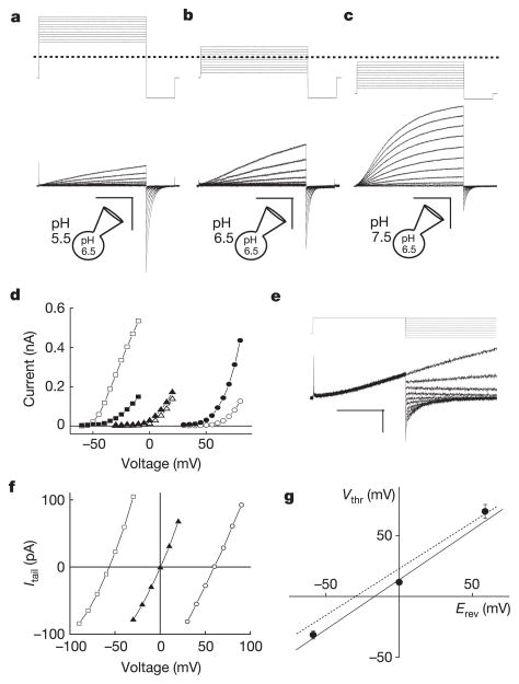
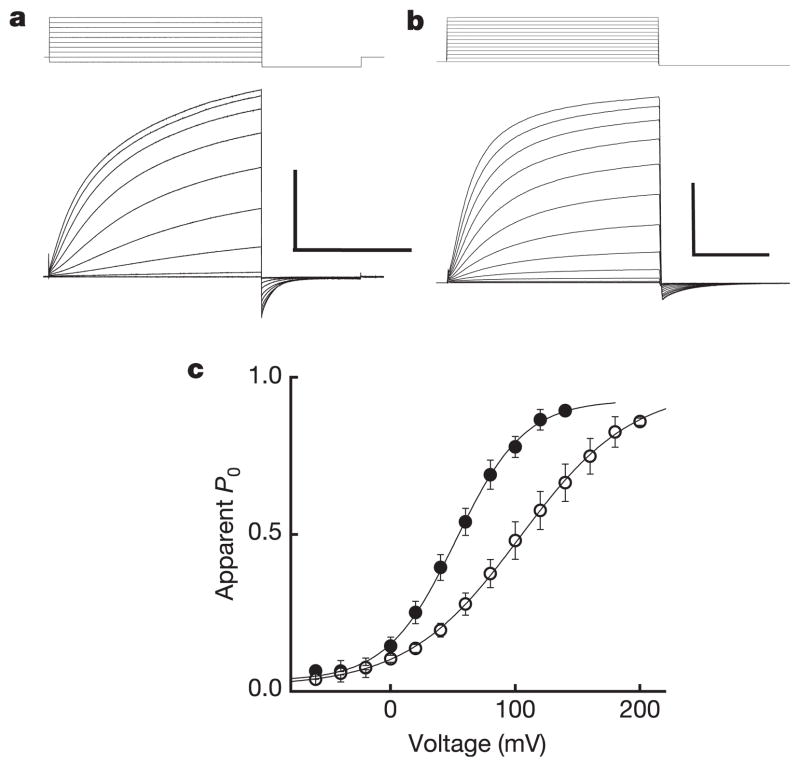
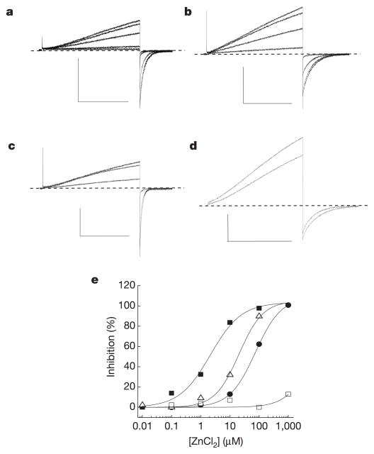
Voltage changes across the cell membrane control the gating of many cation-selective ion channels. Conserved from bacteria to humans, the voltage-gated-ligand superfamily of ion channels are encoded as polypeptide chains of six transmembrane-spanning segments (S1-S6). S1-S4 functions as a self-contained voltage-sensing domain (VSD), in essence a positively charged lever that moves in response to voltage changes. The VSD 'ligand' transmits force via a linker to the S5-S6 pore domain 'receptor', thereby opening or closing the channel. The ascidian VSD protein Ci-VSP gates a phosphatase activity rather than a channel pore, indicating that VSDs function independently of ion channels. Here we describe a mammalian VSD protein (H(V)1) that lacks a discernible pore domain but is sufficient for expression of a voltage-sensitive proton-selective ion channel activity. H(v)1 currents are activated at depolarizing voltages, sensitive to the transmembrane pH gradient, H+-selective, and Zn2+-sensitive. Mutagenesis of H(v)1 identified three arginine residues in S4 that regulate channel gating and two histidine residues that are required for extracellular inhibition of H(v)1 by Zn2+. H(v)1 is expressed in immune tissues and manifests the characteristic properties of native proton conductances (G(vH+)). In phagocytic leukocytes, G(vH+) are required to support the oxidative burst that underlies microbial killing by the innate immune system. The data presented here identify H(v)1 as a long-sought voltage-gated H+ channel and establish H(v)1 as the founding member of a family of mammalian VSD proteins.


Conflict of interest statement

The authors declare no competing financial interests.

Figures

Figure 1
Figure 1. Biophysical properties of expressed Hv1 currents
Depolarizing voltage steps (5 mV increments) were applied to an Hv1-transfected HM1 cell (ac) to elicit outward H+ currents and deactivating inward tail currents (−80 mV). TMA5.5o, TMA6.5o or TMA7.5o (TMA6.5i for all) were used to impose pH gradients indicated in the diagrams. a, ρHi/o+=0.1, Vh = −40 mV (Vstep = +30 mV to +80 mV, scale bar 0.2 nA, 1 s; b, ρHi/o+=1, Vh = −40 mV (Vstep = −30 mV to +20 mV), scale bar 0.1 nA, 1 s; c, ρHi/o+=10, Vh = −70 mV (Vstep = −60 mV to −10 mV), scale bar 0.2 nA, 1 s. d, Hv1 currents at the end of the depolarizing step (Istep, open symbols) and the absolute value of Itail (−80 mV, filled symbols) are plotted as a function of the step voltage. Data shown are from the cell shown in ac. Circles, ρHi/o+=0.1 (Vthr = 70 ± 5.8 mV, n = 3); triangles, ρHi/o+=1.0 (Vthr = 11.7 ± 2.1 mV, n = 6); squares, ρHi/o+=10 (Vthr = −31.7 ± 3.3 mV, n = 3). e, Representative currents for Erev measurement (Vh = −40 mV, Vstep = +40 mV, Vtail = −100 mV to +40 mV). Small Istep (<200 pA) was chosen to minimize Hi+ depletion (note constant outward current level). f, Monoexponential fits of Itail were extrapolated to t = 0 and Erev was estimated from the zero-current intercept of linear fits to the data. Open circles, TMA6.5i, TMA5.5o ( ρHi/o+=0.1); filled triangles, TMA6.5i, TMA6.5o ( ρHi/o+=1); open squares, TMA6.5i, TMA7.5o ( ρHi/o+=10). Average Erev values: Erev = 53.3 ± 1.4 mV, ρHi/o+=0.1, n = 10; Erev = 0.9 ± 0.7 mV, ρHi/o+=1.0, n = 14; Erev = −56.5 ± 2.6 mV, ρHi/o+=10, n = 5. Data represent mean ± s.e.m. from n experiments. g, Vthr is plotted against the Nernst potential for H+ at 24 °C. The data are fitted to Vthr = 0.82 Erev + 13.8 mV (solid line). Data represent mean ± s.e.m. from n = 3–7 experiments. The Vthr versus Erev relationship for native GvH+ (Vthr = 0.79 Erev + 23 mV, dotted line) is shown for comparison.
Figure 2
Figure 2. Hv1 voltage-dependent gating
a, Hv1 currents (−60 mV to +120 mV, Vh = −40 mV, ρHi/o+=10, Na6.5i, Na7.5o) in a representative cell. Scale bar 2 nA, 400 ms. Under symmetrical conditions (TMA6.5, ρHi/o+=1), τact = 715 ± 124 ms (+80 mV), n = 9 and τdeact = 65.0 ± 12.1 ms (−80 mV), n = 7. b, R205A (−60 mV to +180 mV, Vh = −60 mV, ρHi/o+=10, Na6.5i, Na7.5o). Scale bar 2 nA, 10 ms. Note the 40-fold difference in timescale compared to a. c, The ItailV relation (−80 mV, ρHi/o+=10, Na6.5i, Na7.5o) was normalized to the maximum current obtained from a Boltzmann fit to the data for each cell expressing Hv1 (filled circles) or R205A (open circles) to estimate PoV. Data were fitted to a Boltzmann function (solid lines) and normalized to the extrapolated maximum current. Points represent mean ± s.e.m. of normalized data. A comparison of curve fits from individual experiments (Hv1, V0.5 = 58.0 ± 5.6 mV, = 0.90 ± 0.04, n = 6; R205A, V0.5 = 99.5 ± 18.3 mV, = 0.57* ± 0.03, n = 3; *P = 0.03 by Student’s non-paired t-test) indicates that significantly less effective charge is moved in R205A than in Hv1 during channel gating. Data represent mean ± s.e.m. from n experiments.
Figure 3
Figure 3. Mutations in Hv1 reveal residues required for Zn2+ inhibition
Inhibition of Istep (+25 mV to +50 mV) in cells superfused with EGTA-free TMA6.5 or Na6.5 ( ρHi/o+=1) was typically faster than the sampling interval (10 s) and washout was always complete. Average IC50 = 2.2 ± 0.6 μM, nH = 1.0 ± 0.2, n = 4; ρHi/o+=1, + 40 mV, TMA6.5. Data represent mean ± s.e.m. from n experiments. The applied voltages and [Zn2+] for the records shown (Na6.5, ρHi/o+=1) were: a, Hv1, Vstep = +40/Vtail = −80 mV, [Zn2+] = 0, 0.1, 1, 10, 100 μM; b, H140A, +40/−60 mV, [Zn2+] = 0, 1, 10, 100 μM; c, H193A, +30/−80 mV, [Zn2+] = 0, 10, 100 μM; d, H140A/H193A, +50/−20 mV, [Zn2+] = 0, 1 mM. Scale bars: 100 pA, 1 s. e, Representative Zn2+ concentration-response curves for Hv1 (filled squares, IC50 = 1.9 μM, nH = 0.9), H193A (open triangles, IC50 = 17.9 μM, nH = 1.2), H140A (filled circles, IC50 = 74.3 μM, nH = 1.0), and H140A/H193A (open squares, maximum inhibition = 11.1 ± 3.4%, n = 3).

References

    1. Yu FH, Catterall WA. The VGL-chanome: a protein superfamily specialized for electrical signaling and ionic homeostasis. Sci STKE. 2004;2004:re15. - PubMed
    1. Long SB, Campbell EB, Mackinnon R. Crystal structure of a mammalian voltage-dependent Shaker family K+ channel. Science. 2005;309:897–903. - PubMed
    1. Murata Y, Iwasaki H, Sasaki M, Inaba K, Okamura Y. Phosphoinositide phosphatase activity coupled to an intrinsic voltage sensor. Nature. 2005;435:1239–1243. - PubMed
    1. DeCoursey TE. Voltage-gated proton channels and other proton transfer pathways. Physiol Rev. 2003;83:475–579. - PubMed
    1. DeCoursey TE, Morgan D, Cherny VV. The voltage dependence of NADPH oxidase reveals why phagocytes need proton channels. Nature. 2003;422:531–534. - PubMed

Publication types