. 2006 Mar 23;440(7083):551-5.
doi: 10.1038/nature04600.

CHIP-mediated stress recovery by sequential ubiquitination of substrates and Hsp70

Affiliations

CHIP-mediated stress recovery by sequential ubiquitination of substrates and Hsp70

Shu-Bing Qian et al. Nature. .

Abstract

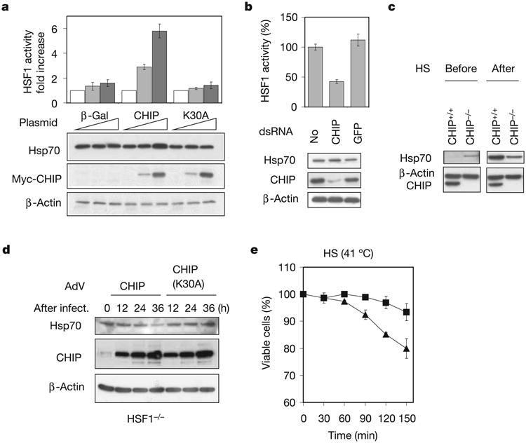
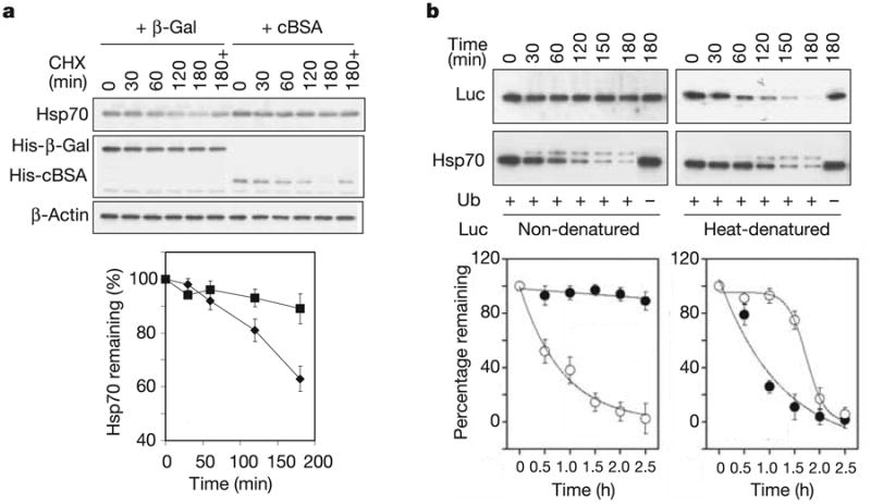
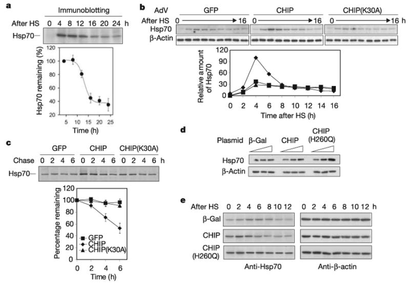
Exposure of cells to various stresses often leads to the induction of a group of proteins called heat shock proteins (HSPs, molecular chaperones). Hsp70 is one of the most highly inducible molecular chaperones, but its expression must be maintained at low levels under physiological conditions to permit constitutive cellular activities to proceed. Heat shock transcription factor 1 (HSF1) is the transcriptional regulator of HSP gene expression, but it remains poorly understood how newly synthesized HSPs return to basal levels when HSF1 activity is attenuated. CHIP (carboxy terminus of Hsp70-binding protein), a dual-function co-chaperone/ubiquitin ligase, targets a broad range of chaperone substrates for proteasomal degradation. Here we show that CHIP not only enhances Hsp70 induction during acute stress but also mediates its turnover during the stress recovery process. Central to this dual-phase regulation is its substrate dependence: CHIP preferentially ubiquitinates chaperone-bound substrates, whereas degradation of Hsp70 by CHIP-dependent targeting to the ubiquitin-proteasome system occurs when misfolded substrates have been depleted. The sequential catalysis of the CHIP-associated chaperone adaptor and its bound substrate provides an elegant mechanism for maintaining homeostasis by tuning chaperone levels appropriately to reflect the status of protein folding within the cytoplasm.


Conflict of interest statement

The authors declare no competing financial interests.

Figures

Figure 1
Figure 1. CHIP regulates Hsp70 availability
a, HEK-293 cells were transfected with plasmids as indicated with 0 μg (white bars), 0.1 μg (grey bars) and 0.5 μg (black bars) per well in a six-well plate. HSF1 activity was measured by dual luciferase reporter assay. Hsp70 levels were determined by immunoblotting. b, HEK-293 cells were transfected with dsRNA as indicated. Both HSF1 activity and Hsp70 levels were determined. c, Immortalized CHIP+/+ and CHIP−/− cell lines were heat shocked for 15 min at 42 °C (HS) and left to recover for 4 h at 37 °C. Cell lysates from before and after heat shock were immunoblotted with Hsp70 antibody. d, HSF1−/− cells were infected with AdV expressing CHIP or CHIP(K30A). Hsp70 levels were determined at different times after infection by immunoblotting of whole cell lysates. e, AdV-infected HSF1−/− cells were incubated at 41 °C for various durations and cell viability was measured. Triangles, AdV/CHIP; squares, AdV/CHIP(K30A). Values are means ± s.d.
Figure 2
Figure 2. Selective effects of CHIP on Hsp70 in vivo and in vitro
a, CHIP−/− cells were infected with AdV expressing GFP, CHIP or CHIP(K30A). Infected cells were incubated for 30 min at 42 °C and left to recover for 6 h at 37 °C to induce Hsp expression. Cycloheximide (CHX) was then added and cell aliquots were collected at the times indicated. Whole cell lysates were immunoblotted with a panel of antibodies. The effects of CHIP on Hsp levels were quantified by subtracting their turnover in the presence of GFP. Squares, Hsp70; diamonds, Hsp90; triangles, Hsc70; circles, Grp78. Values are means ± s.d. b, AdV-infected CHIP−/− cells were radiolabelled with [35S]methionine for 30 min at 3 h after heat shock and chased with unlabelled methionine for up to 8 h. Immunoprecipitations were performed with monoclonal antibody against Hsp70 or Hsc70. c, In vitro ubiquitination assays were performed by incubating equal amounts of Hsc70 and Hsp70 in reactions containing E1, E2 (UbcH5a), CHIP, ubiquitin (Ub) and ATP. Purified 26S proteasomes were included in the reaction mixture for the coupled degradation assay. Levels of Hsc70 (squares) and Hsp70 (diamonds) were determined by immunoblotting and quantified by densitometry. Values are means ± s.d. d, In vitro ubiquitination assays were performed with ubiquitin mutants bearing a single lysine as indicated, and the reaction was stopped after 120 min incubation. WT, wild-type.
Figure 3
Figure 3. Substrate-regulated Hsp70 turnover in vivo and in vitro
a, HEK-293 cells were co-transfected with plasmids expressing CHIP, Flag-Hsp70 and cBSA (squares) orβ-Gal (diamonds). The turnover of Hsp70 was determined with the use of a cycloheximide (CHX) chase followed by immunoblotting. ‘180 + ’ indicates the presence of 20 μM MG132 during the chase. Values are means ± s.d. b, In vitro degradation assays were performed with Hsp70 (open circles) and luciferase (Luc; filled circles) as substrates. For heat-denatured luciferase, luciferase was heated at 43 °C for 10 min with Hsp70 and Hdj2 (ref. 20). Both the luciferase and Hsp70 levels were determined in the same samples by immunoblotting and quantified by densitometry. Values are means ± s.d.
Figure 4
Figure 4. CHIP orchestrates both stress response and recovery processes
a, Mouse fibroblasts were heat shocked (HS) at 42 °C for 10 min, and aliquots were collected every 4 h during recovery at 37 °C. Hsp70 levels were determined by immunoblotting. Values are means ± s.d. b, CHIP−/− cells were infected with AdV expressing GFP (squares), CHIP (diamonds) or CHIP(K30A) (triangles). Infected cells were heat shocked at 42 °C for 10 min, and aliquots were collected every 2 h during recovery at 37 °C. Hsp70 levels were determined by immunoblotting and quantified by densitometry. c, The same AdV-infected CHIP−/− cells as in b were heat shocked at 42 °C for 10 min, and radiolabelled with [35S]methionine for 30 min at 3 h after HS and chased with unlabelled methionine for up to 6 h. Immunoprecipitation was performed with a Hsp70-specific antibody and quantified with a PhosphorImager. Symbols are as in b. Values are means ± s.d. d, HEK-293 cells were transfected with plasmids as indicated with 0, 0.1 and 0.5 μg per well in a six-well plate. Hsp70 levels were determined by immunoblotting. e, Transfected HEK-293 cells were heat shocked 42 °C for 5 min, and aliquots were collected every 2 h during recovery at 37 °C. Hsp70 levels were determined by immunoblotting.

References

    1. Parsell DA, Lindquist S. The function of heat-shock proteins in stress tolerance: degradation and reactivation of damaged proteins. Annu Rev Genet. 1993;27:437–496. - PubMed
    1. Morimoto RI. Cells in stress: transcriptional activation of heat shock genes. Science. 1993;259:1409–1410. - PubMed
    1. Feder JH, Rossi JM, Solomon J, Solomon N, Lindquist S. The consequences of expressing hsp70 in Drosophila cells at normal temperatures. Genes Dev. 1992;6:1402–1413. - PubMed
    1. Volloch VZ, Sherman MY. Oncogenic potential of Hsp72. Oncogene. 1999;18:3648–3651. - PubMed
    1. Wu C. Heat shock transcription factors: structure and regulation. Annu Rev Cell Dev Biol. 1995;11:441–469. - PubMed

Publication types