. 2006 Mar 23:1:8.
doi: 10.1186/1745-6150-1-8.

Origin and evolution of the peroxisomal proteome

Affiliations

Origin and evolution of the peroxisomal proteome

Toni Gabaldón et al. Biol Direct. .

Abstract

Background: Peroxisomes are ubiquitous eukaryotic organelles involved in various oxidative reactions. Their enzymatic content varies between species, but the presence of common protein import and organelle biogenesis systems support a single evolutionary origin. The precise scenario for this origin remains however to be established. The ability of peroxisomes to divide and import proteins post-translationally, just like mitochondria and chloroplasts, supports an endosymbiotic origin. However, this view has been challenged by recent discoveries that mutant, peroxisome-less cells restore peroxisomes upon introduction of the wild-type gene, and that peroxisomes are formed from the Endoplasmic Reticulum. The lack of a peroxisomal genome precludes the use of classical analyses, as those performed with mitochondria or chloroplasts, to settle the debate. We therefore conducted large-scale phylogenetic analyses of the yeast and rat peroxisomal proteomes.

Results: Our results show that most peroxisomal proteins (39-58%) are of eukaryotic origin, comprising all proteins involved in organelle biogenesis or maintenance. A significant fraction (13-18%), consisting mainly of enzymes, has an alpha-proteobacterial origin and appears to be the result of the recruitment of proteins originally targeted to mitochondria. Consistent with the findings that peroxisomes are formed in the Endoplasmic Reticulum, we find that the most universally conserved Peroxisome biogenesis and maintenance proteins are homologous to proteins from the Endoplasmic Reticulum Assisted Decay pathway.

Conclusion: Altogether our results indicate that the peroxisome does not have an endosymbiotic origin and that its proteins were recruited from pools existing within the primitive eukaryote. Moreover the reconstruction of primitive peroxisomal proteomes suggests that ontogenetically as well as phylogenetically, peroxisomes stem from the Endoplasmic Reticulum.

Reviewers: This article was reviewed by Arcady Mushegian, Gáspár Jékely and John Logsdon.

Open peer review: Reviewed by Arcady Mushegian, Gáspar Jékely and John Logsdon. For the full reviews, please go to the Reviewers' comments section.


Figures
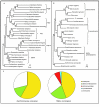
Figure 1
Figure 1
A: Maximum likelihood phylogenetic tree of the CDC48 orthologous group and its paralogs, including PEX1 and PEX6. The crenarchaeon Pyrobaculum aerophilum and euryarchaeon Archaeoglobus fulgidus sequences cluster together, consistent with an ancient eukaryotic origin of this protein family rather than an origin from a horizontal transfer, and they are used as outgroup. PEX1/6, as well as SEC18 and RIX7 appear to have evolved from CDC48, the central protein of the ERAD pathway B: Maximum likelihood phylogenetic tree of the Npy1p orthologous group and its mitochondrial paralogs. This protein family has a single origin in the alpha-proteobacteria. Bootstrap support over 100 replicates of the maximum likelihood tree is shown in all partitions. C: Pie chart showing the relative distribution of peroxisomal proteins according to their phylogenetic origin in yeast (left) and rat (right). Proteins that do have prokaryotic homologs but for which no reliable tree can de constructed, e.g. due to short stretches of homology, are considered "unresolved". For a complete list of the proteins and their origins, see the supplemental material, for their phylogenies see [44].
Figure 2
Figure 2
ERAD and peroxisomal protein import homology. A) Schematic representation of the ERAD (top) and the Pex5 recycling (bottom) pathways. Proteins involved are represented by ovals and rectangles, only those commented in the text are named. Homologous relationships between proteins from the pathways are indicated in color. B) Homology between proteins of the ERAD pathway and proteins involved in protein import to the Peroxisome. Domain organization of the proteins was predicted with SMART [45]. Independent from that, homology between the proteins was determined by profile-to-profile searches using hhsearch [46], based on alignments of orthologous groups of the various proteins. Note that the SEL1 repeat is homologous to the TPR repeat. The location of the two CDC48 N-terminal domains (CDC48_N and CDC48_2) in Pex1 is based on PSI-Blast [47] searches starting with CDC48 proteins and on the structure published for the N-terminal domains of PEX1 [48].
Figure 3
Figure 3
The retargeting of proteins to the peroxisome during evolution. The dashed lines indicate the ancestral cellular location of a peroxisomal protein, the continuous line their current (peroxisomal) location. Some proteins are derived from the alpha-proteobacterial ancestor of the mitochondria, their proteins have been retargeted to the peroxisome concomitant with the transfer of their genes to the nucleus (red, scenario I). Also proteins without a (detectable) alpha-proteobacterial ancestry have been retargeted from the mitochondria (blue, scenario II). Finally, a class of proteins have been retargeted from other compartments of the cell like the Endoplasmic Reticulum (cyan, scenario III).
Figure 4
Figure 4
The N-terminal region of the multiple sequence alignment of several fungal members of the Cit1/2p orthologous group. Amino acids around the signal-peptide cleavage-sites, as predicted by Mitoprot are marked with a rectangle (white arrow indicates the position in the alignment) they correspond to YS (YA in C. tropicalis) that is missing in Cit2p. No mitochondrial localization nor a cleavage-site is predicted for Cit2p consistent with its peroxisomal location.
Figure 5
Figure 5
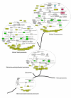
Evolution of the peroxisomal proteome. Biochemical pathways reconstructed according to KEGG and annotations of peroxisomal proteins. For details on the reconstruction of ancestral states see supplemental material. Color code: yellow, eukaryotic origin; green, alpha-proteobacterial origin; red, actinomycetales origin; blue, cyanobacterial origin; white, origin unresolved. Note that the ancestral eukaryotic peroxisomal proteome reconstruction depends on the topology of the eukaryotic tree. If an alternative topology is considered, placing kinetoplastida and viridiplantae together [49], and the plant peroxisomal proteome is taken from the Araperox database [31], then the reconstructed ancestral eukaryotic peroxisomal proteome would be much larger, including all proteins present in the opisthokont proteome except for ANT1, IDP3, FOX3, PEX13 and PEX19.

References

    1. de Duve D. The peroxisome: a new cytoplasmic organelle. Proc R Soc Lond B Biol Sci. 1969;173:71–83. - PubMed
    1. Subramani S. Components involved in peroxisome import, biogenesis, proliferation, turnover, and movement. Physiol Rev. 1998;78:171–188. - PubMed
    1. Latruffe N, Vamecq J. Evolutionary aspects of peroxisomes as cell organelles, and of genes encoding peroxisomal proteins. Biol Cell. 2000;92:389–395. doi: 10.1016/S0248-4900(00)01083-2. - DOI - PubMed
    1. Lazarow PB, Fujiki Y. Biogenesis of peroxisomes. Annu Rev Cell Biol. 1985;1:489–530. doi: 10.1146/annurev.cb.01.110185.002421. - DOI - PubMed
    1. Erdmann R, Kunau WH. A genetic approach to the biogenesis of peroxisomes in the yeast Saccharomyces cerevisiae. Cell Biochem Funct. 1992;10:167–174. doi: 10.1002/cbf.290100306. - DOI - PubMed