

The effect of sustained low-intensity contractions on supraspinal fatigue in human elbow flexor muscles
- PMID: 16556656
- PMCID: PMC1779725
- DOI: 10.1113/jphysiol.2005.103598
The effect of sustained low-intensity contractions on supraspinal fatigue in human elbow flexor muscles
Abstract
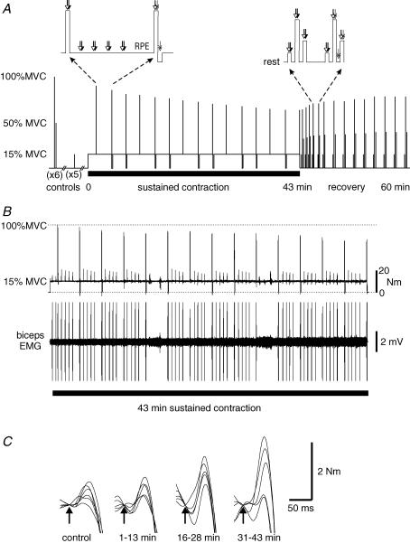
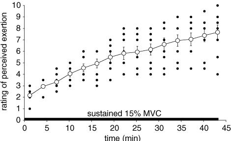
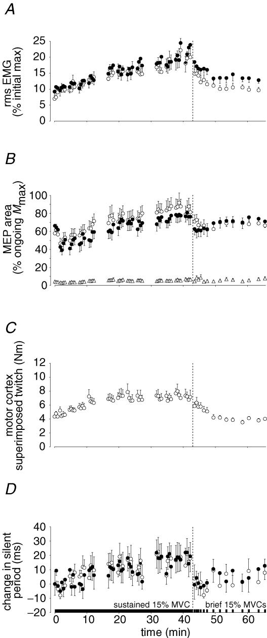
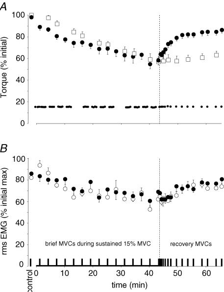
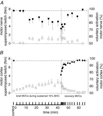
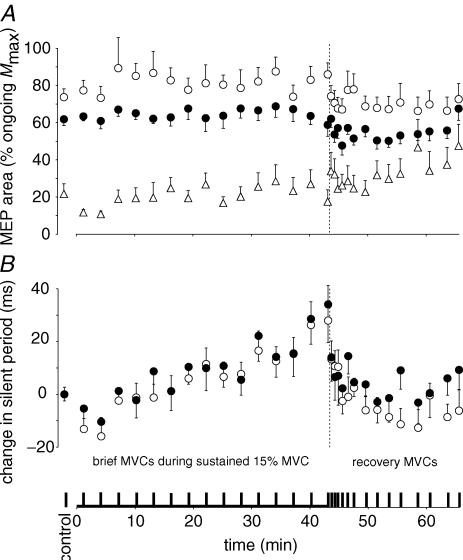
Subjects quickly fatigue when they perform maximal voluntary contractions (MVCs). Much of the loss of force is from processes within muscle (peripheral fatigue) but some occurs because voluntary activation of the muscle declines (central fatigue). The role of central fatigue during submaximal contractions is not clear. This study investigated whether central fatigue developed during prolonged low-force voluntary contractions. Subjects (n=9) held isometric elbow flexions of 15% MVC for 43 min. Voluntary activation was measured during brief MVCs every 3 min. During each MVC, transcranial magnetic stimulation (TMS) was followed by stimulation of either brachial plexus or the motor nerve of biceps brachii. After nerve stimulation, a resting twitch was also evoked before subjects resumed the 15% MVC. Perceived effort, elbow flexion torque and surface EMG from biceps, brachioradialis and triceps were recorded. TMS was also given during the sustained 15% MVC. During the sustained contraction, perceived effort rose from approximately 2 to approximately 8 (out of 10) while ongoing biceps EMG increased from 6.9+/-2.1% to 20.0+/-7.8% of initial maximum. Torque in the brief MVCs and the resting twitch fell to 58.6+/-14.5 and 58.2+/-13.2% of control values, respectively. EMG in the MVCs also fell to 62.2+/-15.3% of initial maximum, and twitches evoked by nerve stimulation and TMS grew progressively. Voluntary activation calculated from these twitches fell from approximately 98% to 71.9+/-38.9 and 76.9+/-18.3%, respectively. The silent period following TMS lengthened both in the brief MVCs (by approximately 40 ms) and in the sustained target contraction (by approximately 18 ms). After the end of the sustained contraction, the silent period recovered immediately, voluntary activation and voluntary EMG recovered over several minutes while MVC torque only returned to approximately 85% baseline. The resting twitch showed no recovery. Thus, as well as fatigue in the muscle, the prolonged low-force contraction produced progressive central fatigue, and some of this impairment of the subjects' ability to drive the muscle maximally was due to suboptimal output from the motor cortex. Although caused by a low-force contraction, both the peripheral and central fatigue impaired the production of maximal voluntary force. While central fatigue can only be demonstrated during MVCs, it may have contributed to the disproportionate increase in perceived effort reported during the prolonged low-force contraction.
Figures
Similar articles
-
Sustained contraction at very low forces produces prominent supraspinal fatigue in human elbow flexor muscles.J Appl Physiol (1985). 2007 Aug;103(2):560-8. doi: 10.1152/japplphysiol.00220.2007. Epub 2007 Apr 26. J Appl Physiol (1985). 2007. PMID: 17463302
-
Supraspinal fatigue does not explain the sex difference in muscle fatigue of maximal contractions.J Appl Physiol (1985). 2006 Oct;101(4):1036-44. doi: 10.1152/japplphysiol.00103.2006. Epub 2006 May 25. J Appl Physiol (1985). 2006. PMID: 16728525
-
Mechanisms of fatigue differ after low- and high-force fatiguing contractions in men and women.Muscle Nerve. 2007 Oct;36(4):515-24. doi: 10.1002/mus.20844. Muscle Nerve. 2007. PMID: 17626289
-
Evidence for a supraspinal contribution to human muscle fatigue.Clin Exp Pharmacol Physiol. 2006 Apr;33(4):400-5. doi: 10.1111/j.1440-1681.2006.04363.x. Clin Exp Pharmacol Physiol. 2006. PMID: 16620309 Review.
-
A comparison of central aspects of fatigue in submaximal and maximal voluntary contractions.J Appl Physiol (1985). 2008 Feb;104(2):542-50. doi: 10.1152/japplphysiol.01053.2007. Epub 2007 Nov 21. J Appl Physiol (1985). 2008. PMID: 18032577 Review.
Cited by
-
Corticospinal responses to sustained locomotor exercises: moving beyond single-joint studies of central fatigue.Sports Med. 2013 Jun;43(6):437-49. doi: 10.1007/s40279-013-0020-6. Sports Med. 2013. PMID: 23456490 Review.
-
Effect of New Zealand Blackcurrant Extract on Isometric Contraction-Induced Fatigue and Recovery: Potential Muscle-Fiber Specific Effects.Sports (Basel). 2020 Oct 15;8(10):135. doi: 10.3390/sports8100135. Sports (Basel). 2020. PMID: 33076273 Free PMC article.
-
The neuromechanical of Beta-band corticomuscular coupling within the human motor system.Front Neurosci. 2024 Aug 15;18:1441002. doi: 10.3389/fnins.2024.1441002. eCollection 2024. Front Neurosci. 2024. PMID: 39211436 Free PMC article. Review.
-
Anodal transcranial direct current stimulation does not influence the neural adjustments associated with fatiguing contractions in a hand muscle.Eur J Appl Physiol. 2019 Mar;119(3):597-609. doi: 10.1007/s00421-018-4027-4. Epub 2018 Nov 13. Eur J Appl Physiol. 2019. PMID: 30421008
-
Exacerbated impairments in neuromuscular function when two bouts of team sport match simulations are separated by 48 h.Exp Physiol. 2023 Nov;108(11):1422-1433. doi: 10.1113/EP091419. Epub 2023 Oct 9. Exp Physiol. 2023. PMID: 37811800 Free PMC article.
References
-
- Adam A, De Luca CJ. Firing rates of motor units in human vastus lateralis muscle during fatiguing isometric contractions. J Appl Physiol. 2005;99:268–280. - PubMed
-
- Allen DG, Kabbara AA, Westerblad H. Muscle fatigue: the role of intracellular calcium stores. Can J Appl Physiol. 2002;27:83–96. - PubMed
-
- Baker AJ, Kostov KG, Miller RG, Weiner MW. Slow force recovery after long-duration exercise: metabolic and activation factors in muscle fatigue. J Appl Physiol. 1993;74:2294–2300. - PubMed
-
- Belanger AY, McComas AJ. Extent of motor unit activation during effort. J Appl Physiol. 1981;51:1131–1135. - PubMed
-
- Bigland-Ritchie B, Cafarelli E, Vollestad NK. Fatigue of submaximal static contractions. Acta Physiol Scand Suppl. 1986a;556:137–148. - PubMed
Publication types
MeSH terms
LinkOut - more resources
Full Text Sources