. 2006 Apr 18;103(16):6208-11.
doi: 10.1073/pnas.0510163103. Epub 2006 Mar 23.

Transgenic Hydra allow in vivo tracking of individual stem cells during morphogenesis

Affiliations

Transgenic Hydra allow in vivo tracking of individual stem cells during morphogenesis

Jörg Wittlieb et al. Proc Natl Acad Sci U S A. .

Abstract

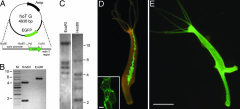
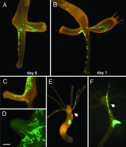
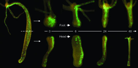
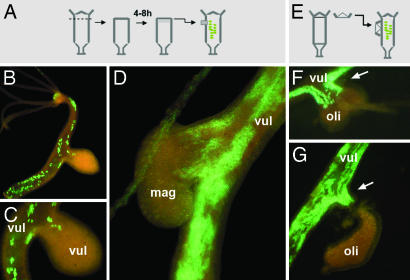
Understanding the evolution of development in large part relies on the study of phylogenetically old organisms. Cnidarians, such as Hydra, have become attractive model organisms for these studies. However, despite long-term efforts, stably transgenic animals could not be generated, severely limiting the functional analysis of genes. Here we report the efficient generation of transgenic Hydra lines by embryo microinjection. One of these transgenic lines expressing EGFP revealed remarkably high motility of individual endodermal epithelial cells during morphogenesis. We expect that transgenic Hydra will become important tools to dissect the molecular mechanisms of development at the base of the Metazoan tree.


Conflict of interest statement

Conflict of interest statement: No conflicts declared.

Figures

Fig. 1.
Fig. 1.
Generation and characterization of transgenic H. vulgaris (AEP) line endo-2. (A) The EGFP expression construct hoT G used to generate transgenic H. vulgaris. (B) hoT G construct digested by EcoRI and HindIII showing one band of ≈5 kb and two bands of 4 and 1.5 kb; M, size marker. (C) Southern blot hybridization. DNA from H. vulgaris (AEP) endo-2 line was hybridized with a probe specific for EGFP. Lane 1, EcoRI-digested DNA from transgenic polyps; lane 2, HindIII-digested DNA from transgenic polyps. The differences in fragment size between the restricted genomic DNA and the hoT G vector (shown in B) indicate stable integration of the vector in the Hydra genome. (D) Transgenic endo-2 polyp with a mosaic expression of EGFP in some of its endodermal epithelial cells. (Inset) A patch of EGFP+ endodermal epithelial cells visualized by confocal microscopy. (Scale bar, 20 μm.) (E) Transgenic endo-2 polyp expressing EGFP in all of its endodermal epithelial cells. (Scale bar, 1 mm.)
Fig. 2.
Fig. 2.
Individual motility of EGFP-expressing endodermal epithelial cells. (A–D) EGFP+ cells reveal high motility of endodermal epithelial cells during budding. (A–C) Buds recruit EGFP+ endodermal epithelial cells even from distant parts of the parent polyp. (D) Confocal image of evaginating bud showing change in epithelial cell shape during budding. (Scale bar, 80 μm.) (E and F) Homotopic transplantation experiments confirm the motility of individual epithelial cells. Small tissue pieces containing EGFP-expressing cells were transplanted homotopically into the gastric region of a nontransgenic recipient. (E) EGFP-positive epithelial cells migrated away from the transplant 5 days after transplantation; arrow points to emigrated endodermal epithelial cells in the tentacle. (F) String of EGFP-positive cells 8 days after transplantation produced as a result of cell migration and oriented cell divisions; arrow points to emigrated endodermal epithelial cells.
Fig. 3.
Fig. 3.
Head and foot regeneration occurs via morphallaxis in the absence of local cell proliferation with no evidence for a piling up of endodermal epithelial cells. Numbers indicate hours after amputation.
Fig. 4.
Fig. 4.
Motility of endodermal cells toward developmental signals from regenerating tissue. (A and E) Experimental procedure. (B and C) Lateral transplantation of regenerating H. vulgaris tissue recruits EGFP+ cells to form a secondary axis. (D) Regenerating tissue from H. magnipapillata induces secondary axis formation and recruits H. vulgaris EGFP+ cells. (F and G) In hypostome-contact grafts, heads from H. oligactis induce H. vulgaris to form a secondary axis by recruiting EGFP+ H. vulgaris endodermal epithelial cells. Arrow points to H. vulgaris EGFP+ endodermal cells, which begin to form a secondary axis. mag, H. magnipapillata; oli, H. oligactis; vul, H. vulgaris.

Comment in

  • Trembley's polyps go transgenic.
    Steele RE. Steele RE. Proc Natl Acad Sci U S A. 2006 Apr 25;103(17):6415-6. doi: 10.1073/pnas.0601983103. Epub 2006 Apr 17. Proc Natl Acad Sci U S A. 2006. PMID: 16618934 Free PMC article. No abstract available.

References

    1. Orr J. C., Fabry V. J., Aumont O., Bopp L., Doney S. C., Feely R. A., Gnanadesikan A., Gruber N., Ishida A., Joos F., et al. Nature. 2005;437:681–686. - PubMed
    1. Gierer A. Sci. Am. 1974;231:44–54. - PubMed
    1. Bosch T. C. G., Fujisawa T. BioEssays. 2001;23:420–427. - PubMed
    1. Galliot B., Schmid V. Int. J. Dev. Biol. 2002;46:39–48. - PubMed
    1. Miller D. J., Ball E. E., Technau U. Trends Genet. 2005;21:536–539. - PubMed

Publication types

Substances