. 2006 Apr 28;98(8):1032-9.
doi: 10.1161/01.RES.0000218782.52610.dc. Epub 2006 Mar 23.

Essential role of Smad3 in angiotensin II-induced vascular fibrosis

Affiliations

Essential role of Smad3 in angiotensin II-induced vascular fibrosis

Wansheng Wang et al. Circ Res. .

Abstract

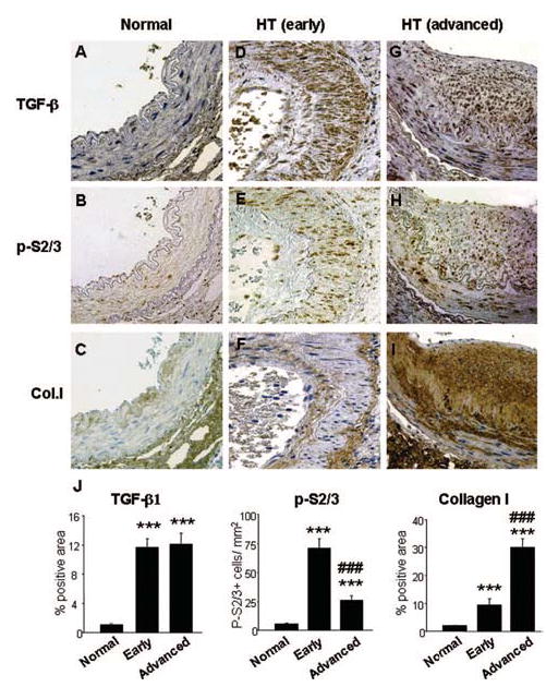
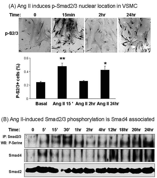
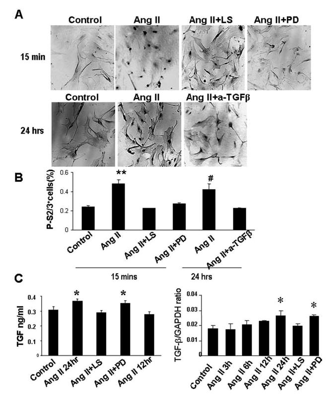
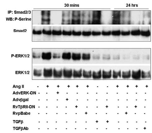
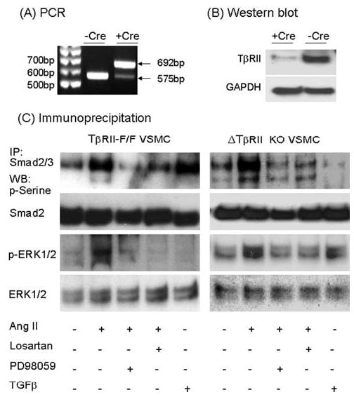
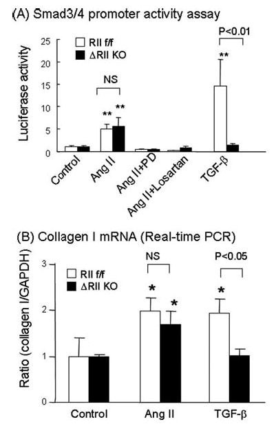
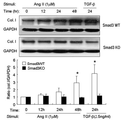
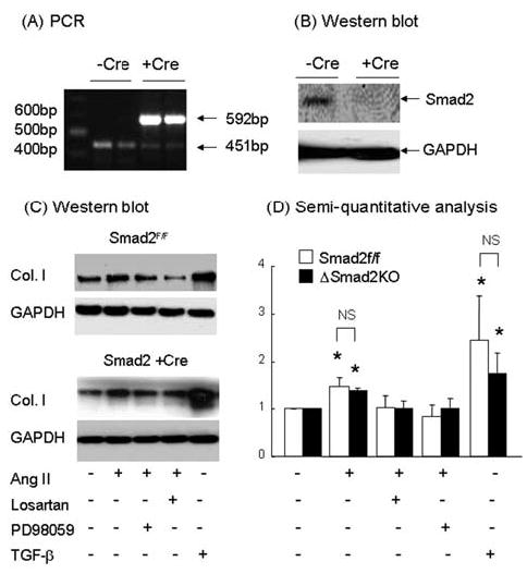
Angiotensin II (Ang II) plays a pivotal role in vascular fibrosis, which leads to serious complications in hypertension and diabetes. However, the underlying signaling mechanisms are largely unclear. In hypertensive patients, we found that arteriosclerosis was associated with the activation of Smad2/3. This observation was further investigated in vitro by stimulating mouse primary aorta vascular smooth muscle cells (VSMCs) with Ang II. There were several novel findings. First, Ang II was able to activate an early Smad signaling pathway directly at 15 to 30 minutes. This was extracellular signal-regulated kinase 1/2 (ERK1/2) mitogen-activated protein kinase (MAPK) dependent but transforming growth factor-beta (TGF-beta) independent because Ang II-induced Smad signaling was blocked by addition of ERK1/2 inhibitor and by dominant-negative (DN) ERK1/2 but not by DN-TGF-beta receptor II (TbetaRII) or conditional deletion of TbetaRII. Second, Ang II was also able to activate the late Smad2/3 signaling pathway at 24 hours, which was TGF-beta dependent because it was blocked by the anti-TGF-beta antibody and DN-TbetaRII. Finally, activation of Smad3 but not Smad2 was a key and necessary mechanism of Ang II-induced vascular fibrosis because Ang II induced Smad3/4 promoter activities and collagen matrix expression was abolished in VSMCs null for Smad3 but not Smad2. Thus, we concluded that Ang II induces vascular fibrosis via both TGF-beta-dependent and ERK1/2 MAPK-dependent Smad signaling pathways. Activation of Smad3 but not Smad2 is a key mechanism by which Ang II mediates arteriosclerosis.


Figures

Figure 1
Figure 1
Immunohistochemistry shows that Smad2/3 is activated in human hypertensive arteriopathy. A through C, Serial sections of a normal human renal artery obtained from paratumor kidney tissues show that there are low levels of TGF-β, p-Smad2/3, and collagen I expression by VSMCs. D through F, Serial sections of a renal artery with the early stage of vascular fibrosis (media:intima >1.0) obtained from a patient with hypertension. Marked activation of p-Smad2/3 is noted in most medial VSMCs (E), which is associated with marked upregulation of TGF-β1 and some collagen I deposition (D and F). G through I, Serial sections of a renal artery with advanced arteriosclerosis (media:intima <1.0) obtained from a patient with hypertension. Note that activation of vascular p-Smad2/3 (H) is associated with upregulation of TGF-β1 and the development of marked accumulation of collagen I in the area of intimal thickening (G and I). J, Quantitative analysis: The results were expressed as mean±SEM. ***P<0.001 compared with normal; ###P<0.001 compared with the early stage of arteriosclerosis. Sections are counterstained with hematoxilin except B, E, and H. Magnification ×200.
Figure 2
Figure 2
Ang II activates Smad2/3 in VSMCs in a biphasic manner. A, Immunohistochemistry shows that Ang II induces early (15 minutes) and a late (24 hours) p-Smad2/3 nuclear translocation (brown nuclei) in VSMCs. B, Immunoprecipitation shows that Ang II induced an early (5 minutes to 1 hour) and a late (18 to 24 hours) p-Smad2/3 in VSMCs, which is Smad4 associated. Data represent three independent experiments.
Figure 3
Figure 3
Signaling mechanisms of Ang II induce the early and late Smad activation in VSMCs shown by immunohistochemistry. A, Immunohistochemistry shows that Ang II (1 μmol/L)–induced early p-Smad2/3 nuclear translocation (brown nuclei) at 15 minutes is blocked by addition of losartan (1 μmol/L) and PD98059 (20 μmol/L). Ang II (1 μmol/L)–induced late p-Smad2/3 nuclear translocation is prevented by addition of a neutralizing TGF-β Ab (10 μg/mL). B, Quantitative analysis. Each bar represents the mean±SEM for three independent experiments. **P<0.01 compared with the results from basal, Ang II + losartan, and Ang II + PD98059; #P<0.05 compared with Ang II + neutralizing TGF-β Ab. Magnifications ×250. C, ELISA (left) and real-time quantitative PCR (right) demonstrated that Ang II induced significant TGF-β expression at 24 hours. The increase of TGF-β expression by Ang II is AT1 dependent but not ERK1/2 dependent. Each bar represents the mean±SEM for three independent experiments. *P<0.05 compared with the control.
Figure 4
Figure 4
Signaling mechanisms of Ang II induce the early and late Smad activation in VSMCs shown by immunoprecipitation (IP). Immunoprecipitation shows that Ang II (1 μmol/L)–induced early phosphorylation (p) of Smad2/3 at 30 minutes in VSMCs is associated with the activation of p-ERK1/2 MAPK, which is blocked by overexpressing Adv-ERK-DN (moi of 50) but not Rv-TβRII-DN, whereas Ang II–induced late activation of p-Smad2/3 at 24 hours is inhibited by overexpressing Rv-TβRII-DN and by neutralizing TGF-β Ab (10 μg/mL). The specificity of Adv-ERK-DN in inhibition of p-ERK1/2 and p-Smad2/3 is confirmed by the inability of a control Adv-β-gal to block activation of both ERK1/2 and Smad2/3. The specificity of Rv-TβRII-DN is also confirmed by a control Rv-pBabe vector and by the ability of Rv-TβRII-DN to block TGF-β (2.5 ng/mL)–induced p-Smad2/3 at 30 minutes. Results represent three independent experiments.
Figure 5
Figure 5
Conditional deletion of TβRII cannot prevent early Ang II–induced Smad2/3 phosphorylation in VSMCs, which is blocked by an ERK1/2 MAPK inhibitor. A, TβRIIf/f VSMCs were treated with Cre-expressing adenovirus (moi of 50) as described in the Methods. PCR shows that Cre recombination substantially deletes the floxed TβRII gene (575 bp) as demonstrated by a high level of the deleted allele (692 bp) (primers sequences are: 1: 5′-TATGGACTGGCTGCTTTTGTATTC; 2: 5′-TGGGGATA-GAGGTAGAAAGACATA; 3: 5′-TATTGGGTGTGGTTGTGGACTTTA). B, Western blot analysis shows that infection of Adv-Cre causes a substantial lose of TβRII protein in TβRIIf/f VSMCs. C, Immunoprecipitation demonstrates that compared with the TβRIIf/f VSMC (left panel), conditional deletion of TβRII cannot prevent Ang II–induced p-ERK1/2 and p-Smad2/3 in VSMCs. In contrast, addition of losartan (1 μmol/L) and PD 98059 (20 μmol/L) is able to block Ang II–induced p-ERK1/2 and p-Smad2/3 in both TβRIIf/f and conditional TβRII KO VSMCs. Effect of conditional deletion of TβRII on TGF-β– dependent Smad signaling in VSMCs is confirmed by the finding that conditional TβRII KO VSMCs are protected against TGF-β (2.5 ng/mL)–induced p-Smad2/3. Data represent three independent experiments.
Figure 6
Figure 6
Functional role of TGF-β–independent ERK1/2 MAPK-dependent Smad signaling pathway in Ang II–induced Smad3/4 promoter activities and collagen I mRNA expression. A, Both TβRIIf/f and conditional KO VSMCs were transiently transfected with p(CAGA)12-Luc and pCMV-β-gal, followed by treatment with Ang II (1 μmol/L) in the presence of PD98059 (20 μmol/L), losartan (1 μmol/L), or TGF-β (1 ng/ ml) for 6 hours. The activity of luciferase and β-gal was measured, and results were expressed as the ratio of luciferase/β-gal (mean±SEM). Results show that conditional deletion of TβRII does not prevent Ang II–induced Smad3/4 promoter activities that are completely blocked by addition of losartan and PD98059. In contrast, conditional deletion of TβRII abrogates TGF-β–induced Smad3/4 promoter activities. B, Real-time PCR shows that conditional deletion of TβRII in VSMCs does not prevent Ang II–induced but is able to block TGF-β–induced collagen I mRNA expression. Results represent three independent experiments. *P<0.05; **P<0.01 compared with the normal control or as indicated. ns indicates not significant.
Figure 7
Figure 7
Essential role for Smad3 in Ang II–induced collagen matrix production. VSMCs were isolated from Smad3 WT and KO mice and stimulated with Ang II (1 μmol/L) for different time as described in the Methods. TGF-β (2.5 ng/mL) was used as a positive control. Western blot analysis shows that Ang II induces significant collagen I expression at 48 hours in Smad3 WT VSMCs, which is completely abrogated in Smad3 KO VSMCs. A similar result is seen in TGF-β stimulation. Results represent three independent experiments. *P<0.05 compared with time-matched Smad3 KO VSMCs.
Figure 8
Figure 8
Smad2 is not necessary for Ang II–induced collagen type I expression. A, Smad2f/f VSMC was treated with Cre-adenovirus as described in the Methods. PCR shows that floxed Smad2 gene is clearly identified in Smad2f/f VSMC without Adv-Cre treatment (451 bp), which is substantially deleted by Cre recombination as identified by the deleted allele (592 bp; primers sequences are: 1: 5′-TTCCATCATCCTTCATGCAAT, 2: 5′-CTTGTGGCAAATGCCCTTAT, 3: 5′- 5′-GACCAAGGCGAAA-GGAAACT). B, Western blot analysis shows that addition of Adv-Cre causes a loss of Smad2 at the protein level. C, Growth-arrested Smad2f/f and conditional Smad2 KO VSMCs were treated with Ang II (1 μmol/L) in the presence of PD98059 (20 μmol/L), losartan (1 μmol/L), or positive control TGF-β (1 ng/mL) for 24 hours. Western blot analysis shows that Ang II as well as TGF-β is able to induce collagen I protein expression in Smad2f/f VSMCs. However, conditional deletion of Smad2 does not prevent Ang II– and TGF-β–induced collagen I expression in VSMCs. In contrast, addition of losartan and PD98059 block Ang II–induced collagen I expression in both Smad2f/f and conditional KO VSMCs. D, Semiquantitative analysis. Each bar represents the mean±SEM from three independent experiments. *P<0.05 compared with control and treatment with losartan and PD98059. ns indicates not significant.

Comment in

References

    1. Ford CM, Li S, Pickering JG, Itoh H, Mukoyama M, Pratt RE, Gibbons GH, Dzau VJ. Angiotensin II stimulates collagen synthesis in human vascular smooth muscle cells. Involvement of the AT(1) receptor, transforming growth factor-beta, and tyrosine phosphorylation. Arterioscler Thromb Vasc Biol. 1999;19:1843–1851. - PubMed
    1. Miao CY, Tao X, Gong K, Zhang SH, Chu ZX, Su DF. Arterial remodeling in chronic sinoaortic-denervated rats. J Cardiovasc Pharmacol. 2001;37:6–15. - PubMed
    1. Lombardi DM, Viswanathan M, Vio CP, Saavedra JM, Schwartz SM, Johnson RJ. Renal and vascular injury induced by exogenous angiotensin II is AT1 receptor-dependent. Nephron. 2001;87:66–74. - PubMed
    1. Hayashi T, Sohmiya K, Ukimura A, Endoh S, Mori T, Shimomura H, Okabe M, Terasaki F, Kitaura Y. Angiotensin II receptor blockade prevents microangiopathy and preserves diastolic function in the diabetic rat heart. Heart. 2003;89:1236–1242. - PMC - PubMed
    1. Kakinuma Y, Kawamura T, Bills T, Yoshioka T, Ichikawa I, Fogo A. Blood pressure-independent effect of angiotensin inhibition on vascular lesions of chronic renal failure. Kidney Int. 1992;42:46–55. - PubMed

Publication types

MeSH terms