. 2006 Apr 28;98(8):1081-8.
doi: 10.1161/01.RES.0000218493.09370.8e. Epub 2006 Mar 23.

A specific pattern of phosphodiesterases controls the cAMP signals generated by different Gs-coupled receptors in adult rat ventricular myocytes

Affiliations

A specific pattern of phosphodiesterases controls the cAMP signals generated by different Gs-coupled receptors in adult rat ventricular myocytes

Francesca Rochais et al. Circ Res. .

Abstract

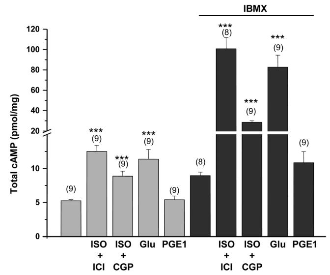
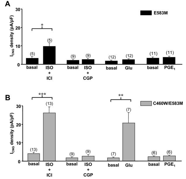
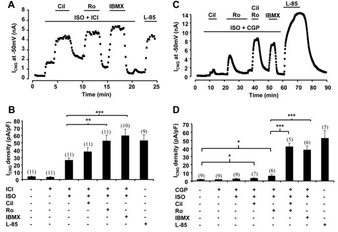
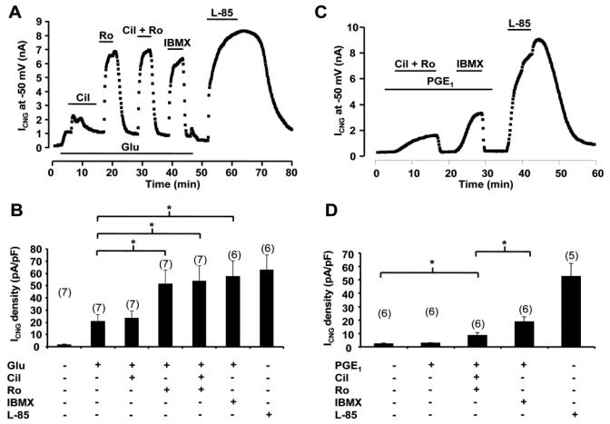
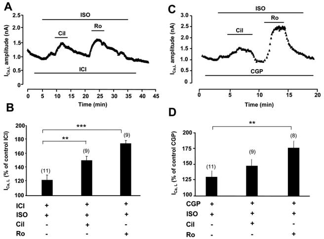
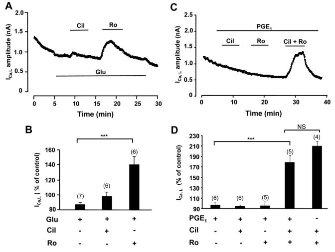
Compartmentation of cAMP is thought to generate the specificity of Gs-coupled receptor action in cardiac myocytes, with phosphodiesterases (PDEs) playing a major role in this process by preventing cAMP diffusion. We tested this hypothesis in adult rat ventricular myocytes by characterizing PDEs involved in the regulation of cAMP signals and L-type Ca2+ current (I(Ca,L)) on stimulation with beta1-adrenergic receptors (beta1-ARs), beta2-ARs, glucagon receptors (Glu-Rs) and prostaglandin E1 receptors (PGE1-Rs). All receptors but PGE1-R increased total cAMP, and inhibition of PDEs with 3-isobutyl-1-methylxanthine strongly potentiated these responses. When monitored in single cells by high-affinity cyclic nucleotide-gated (CNG) channels, stimulation of beta1-AR and Glu-R increased cAMP, whereas beta2-AR and PGE1-R had no detectable effect. Selective inhibition of PDE3 by cilostamide and PDE4 by Ro 20-1724 potentiated beta1-AR cAMP signals, whereas Glu-R cAMP was augmented only by PD4 inhibition. PGE1-R and beta2-AR generated substantial cAMP increases only when PDE3 and PDE4 were blocked. For all receptors except PGE1-R, the measurements of I(Ca,L) closely matched the ones obtained with CNG channels. Indeed, PDE3 and PDE4 controlled beta1-AR and beta2-AR regulation of I(Ca,L), whereas only PDE4 controlled Glu-R regulation of I(Ca,L) thus demonstrating that receptor-PDE coupling has functional implications downstream of cAMP. PGE1 had no effect on I(Ca,L) even after blockade of PDE3 or PDE4, suggesting that other mechanisms prevent cAMP produced by PGE1 to diffuse to L-type Ca2+ channels. These results identify specific functional coupling of individual PDE families to Gs-coupled receptors as a major mechanism enabling cardiac cells to generate heterogeneous cAMP signals in response to different hormones.


Figures

Figure 1
Figure 1
Relative PDE activities in ARVMs. PDE2 activity was determined as the fraction of total hydrolytic activity inhibited by 10 μmol/L EHNA; PDE3 as the fraction inhibited by 1 μmol/L cilostamide; and PDE4 activity as the fraction inhibited by 1 μmol/L rolipram. To some instances, rolipram was replaced by RS25344 (1 μmol/L) or piclamilast (1 μmol/L) with identical results. The bars represent the mean±s.e.m of the number of independent experiments indicated.
Figure 2
Figure 2
Effect of various stimuli linked to adenylyl cyclase activation on total cAMP content in freshly isolated ARVMs. Where necessary, the β1-AR antagonist CGP 20712A (CGP, 1 μmol/L), the β2-AR antagonist ICI 118551 (ICI, 1 μmol/L) and IBMX (100 μmol/L) were added 15 min before the 3 min stimulation with ISO (5 μmol/L), glucagon (Glu, 1 μmol/L) or PGE1 (1 μmol/L). The bars show the means±s.e.m. of the number of experiments indicated near the bars. Statistically significant difference in cAMP content between basal and agonists alones or between IBMX and agonists with IBMX are indicated as ***, p<0.001.
Figure 3
Figure 3
Subsarcolemmal cAMP signals reported by CNG channels upon activation of distinct GsPCRs in ARVMs. CNG current (ICNG) density from recombinant E583M (A) and C460W/E583M (B) CNGA2 channels was measured by the whole-cell patch-clamp technique in rat ventricular myocytes 24 h after isolation. Activation of β1-AR was achieved by application of isoprenaline (ISO, 5 μmol/L) in the presence of the ICI 118551 (ICI, 1 μmol/L). Activation of β2-AR was done by application of ISO (5 μmol/L) in combination with CGP 20712A (CGP, 1 μmol/L). Activation of Glu-R (C) and PGE1-R (D) was done by application of glucagon (Glu, 1 μmol/L) and PGE1 (1 μmol/L). Statistically significant differences are indicated as *, p<0.05; **, p<0.01 and ***, p<0.001.
Figure 4
Figure 4
PDE regulation of cAMP signals from β1-AR and β2-AR. A and C, Time course of ICNG in ARVMs expressing C460W/E583M CNGA2. The cells were first superfused with control external Ringer and then challenged with the drugs during the periods indicated by the solid lines. B and D, Summary of the results obtained in a series of experiments as in A and C, respectively. Specific activation of β1-AR and β2-AR as in Fig. 1. Selective PDE3 inhibition by cilostamide (Cil, 1 μmol/L) or selective PDE4 inhibition by Ro 20-1724 (Ro, 10 μmol/L) strongly potentiated cAMP triggered by β1-AR. Upon activation of β2-AR, robust cAMP accumulation necessitated concomitant PDE3 and PDE4 blockade. Experiment was ended with application of 100 μmol/L L-85. .Statistically significant differences as in figure 3.
Figure 5
Figure 5
PDE regulation of cAMP signals from Glu-R and PGE1-R. A and C, Time course of ICNG in ARVMs expressing C460W/E583M CNGA2. B and D, Summary of the results obtained in a series of experiments as in A and C, respectively. A and B, Cil (1 μmol/L) exerted a transient potentiation of ICNG previously enhanced by glucagon (Glu, 1 μmol/L), while Ro (10 μmol/L) exerted a major and sustained cAMP accumulation. C and D, The cAMP response to PGE1 was revealed at the membrane by simultaneous blockade of PDE3 and PDE4 and further increased by application of IBMX. Statistically significant differences are indicated as *, p<0.05.
Figure 6
Figure 6
Regulation of ICa,L by β-AR and PDEs. A and C, Time course of ICa,L in ARVMs cultured for 24h. The cells were first superfused with control external Ringer and then challenged with the drugs during the periods indicated by the solid lines. ISO was used at 1 nmol/L in A and B, and at 5 μmol/L in C and D; all other drugs as in previous figures. B and D, Summary of the results obtained in a series of experiments as in A and C, respectively. Statistically significant differences indicated as in previous figures.
Figure 7
Figure 7
Regulation of ICa,L by Glu-R, PGE1-R and PDEs. A and C, Time course of ICa,L in ARVMs cultured for 24h. Glucagon (Glu) was used at 1 nmol/L in A and B; all other drugs as in previous figures. B and D, Summary of the results obtained in a series of experiments as in A and C, respectively. Statistically significant differences indicated as in previous figures. NS, non significant.

References

    1. Xiao RP, Lakatta EG. Beta 1-adrenoceptor stimulation and beta 2-adrenoceptor stimulation differ in their effects on contraction, cytosolic Ca2+, and Ca2+ current in single rat ventricular cells. Circ Res. 1993;73:286–300. - PubMed
    1. Bartel S, Krause EG, Wallukat G, Karczewski P. New insights into beta2-adrenoceptor signaling in the adult rat heart. Cardiovasc Res. 2003;57:694–703. - PubMed
    1. Kuznetsov V, Pak E, Robinson RB, Steinberg SF. Beta 2-adrenergic receptor actions in neonatal and adult rat ventricular myocytes. Circ Res. 1995;76:40–52. - PubMed
    1. Farah AE. Glucagon and the circulation. Pharmacol Rev. 1983;35:181–217. - PubMed
    1. Hayes JS, Brunton LL, Brown JH, Reese JB, Mayer SE. Hormonally specific expression of cardiac protein kinase activity. Proc Natl Acad Sci U S A. 1979;76:1570–1574. - PMC - PubMed

Publication types